Edible Cholera Vaccine – Made of Powdered Rice – Proves Safe in Phase 1 Human Trials
MucoRice Cholera Vaccine

Researchers at the University of Tokyo have announced the successful results of the Phase 1 clinical trial of a new type of vaccine to protect against cholera and travelers’ diarrhea. The MucoRice-CTB vaccine is grown in rice plants and stimulates immunity through the mucosal membranes of the intestines. The vaccine can be stored and transported without refrigeration and does not need needles; it is simply mixed with liquid and drunk. Credit: Image by Dr. Hiroshi Kiyono, CC BY 4.0

Study points towards role of gut microbiome in vaccine effectiveness.

A new vaccine to protect against deadly cholera has been made by grinding up genetically modified grains of rice. The first human trial has shown no obvious side effects and a good immune response. Researchers based at the University of Tokyo and Chiba University have published the peer-reviewed results of the Phase 1 clinical trial of the vaccine, named MucoRice-CTB, in The Lancet Microbe.

Vaccine manufacturing has made enormous strides in 2020, spurred on by COVID-19. However, the complexity of mRNA-based SARS-CoV-2 vaccines has highlighted the value of inoculations that can be made, transported and stored cheaply and without refrigeration.

The MucoRice-CTB vaccine is stable at room temperature from start to finish.

“I’m very optimistic for the future of our MucoRice-CTB vaccine, especially because of the dose escalation results. Participants responded to the vaccine at the low, medium and high doses, with the largest immune response at the highest dose,” said Professor Hiroshi Kiyono, D.D.S., Ph.D., from the Institute of Medical Science at the University of Tokyo who leads the MucoRice project. Dr. Kiyono is also a faculty member at Chiba University in Japan and the University of California, San Diego, in the U.S.

MucoRice-CBT Vaccine Trial

Researchers at the University of Tokyo have announced the successful results of the Phase 1 clinical trial of a new type of vaccine to protect against cholera and travelers’ diarrhea. The cartoon shows a simplified summary of the MucoRice-CTB vaccine trial. Credit: Image by Dr. Hiroshi Kiyono, CC BY 4.0

Thirty volunteers received a placebo and groups of 10 volunteers received a total of four doses spaced every two weeks of either 3 milligrams (mg), 6 mg or 18 mg each of the vaccine. Tests two and four months after receiving the last dose revealed that volunteers who responded to the vaccine had IgA and IgG antibodies — two types of proteins the immune system produces to fight infections — specific to cholera toxin B (CTB). Participants who received a higher dose of vaccine were more likely to have CTB-specific antibodies.

An independent review board found no evidence of significant side effects.

Growing a new type of vaccine Vibrio cholerae bacteria is spread most often by drinking water contaminated with sewage. Without medical attention, cholera can kill in mere hours due to diarrhea with severe dehydration. Cholera infects 1.3 million to 4 million people and causes 21,000 to 143,000 deaths each year, according to the World Health Organization.

There are four modern needle-free cholera vaccines, all of which are given as drops on the tongue, but require cold storage and are made from whole killed or live-attenuated (weakened) cholera cells.

The new cholera vaccine grows in genetically modified Japanese short-grain rice plants that produce a nontoxic portion of CTB that can be recognized by the immune system. CTB is similar in structure to a toxin made by some types of disease-causing E. coli bacteria, so cholera vaccines often provide cross protection against travelers’ diarrhea.

Researchers grow the rice plants in a purpose-built, indoor hydroponic farm that meets WHO good manufacturing practice standards for medicines, which ensures that the vaccine remains uncontaminated and that the plants are isolated from the natural environment.

The plants produce the CTB subunit in their seeds, the edible grains of rice, and store the antigens in droplets called protein bodies with membranes made of fat.

“The rice protein bodies behave like a natural capsule to deliver the antigen to the gut immune system,” said Dr. Kiyono.

Other medicines have been grown in plants, most often in the leaves — including treatments for Ebola, lymphoma and flu — but the drugs have to be extracted and purified before being used. The grain-based aspect of the MucoRice system avoids those extra steps, the need for cold storage, and protects the antigens as they travel through the harsh acid of the stomach.

When the plants are mature, the rice is harvested and ground into a fine powder, then sealed in aluminum packets for storage. When people are ready to be vaccinated, the powder is mixed with about 90 milliliters (1/3 U.S. cup) of liquid and then drunk. Researchers have only tested the vaccine using saline (a salt solution equivalent to body fluids), but they expect it would work equally well with plain water.

Immunity through the gut is strong, but complicated by the microbiome

“The beautiful part of our vaccine is that it wisely uses the body’s mucosal immune system through the gut for the induction of antigen-specific antibodies,” said Dr. Kiyono.

MucoRice-CTB enters the body through intestinal mucosal membranes, mimicking a natural way of encountering and responding to germs. Stimulating the mucosal immune system produces two classes of antibodies that identify germs and target them for removal, IgG and IgA. Vaccines that are injected under the skin or into a muscle generally increase only IgG, not IgA, antibodies.

Volunteers who responded to MucoRice-CTB had their highest blood levels of antigen-specific IgG and IgA after eight to 16 weeks.

However, 11 of the 30 volunteers who received the vaccine showed low or no measurable immune response. All study volunteers reported never traveling outside of Japan, so it is unlikely that they had any previous exposure or natural immunity to V. cholerae or pathogenic E. coli.

“When we saw those data about the 11 low and nonresponders, we thought maybe gut microflora have an influence on the outcome of the immune response,” Dr. Kiyono recalled.

The microflora or microbiome is the community of microorganisms that live in our bodies and either benefit us or are harmless. It is well accepted that the microflora of the digestive system influence health and immunity, but scientists are just beginning to understand the precise mechanisms of the relationship.

Extensive genetic analysis of all volunteers’ fecal samples identified the thousands of bacterial species living in volunteers’ intestines.

“In simplified terms, high responders had more diversified microflora, and in the low-responder group, diversity was much narrower,” said Dr. Kiyono.

Researchers cautioned that the small size of the Phase 1 study — giving the vaccine to only 30 healthy Japanese male volunteers — means the relevance and prevalence of nonresponders is still unclear and that the total difference in microflora diversity was subtle. However, the results do hint at the larger role of microflora in vaccine effectiveness.

“It’s all speculation right now, but maybe higher microflora diversity creates a better situation for strong immune response against oral vaccine,” said Dr. Kiyono.

The link between the gut microbiome and vaccine effectiveness has been previously revealed by the unfortunate fact that most vaccines are developed in industrialized nations and some are then less effective when delivered in developing countries. Mucosal vaccines, including oral vaccines against polio and cholera, seem especially prone to this disparity. Most scientific theories to explain the phenomenon focus on chronic intestinal inflammation linked to poor sanitation. (https://doi.org/10.1186/1741-7007-8-129)

“Probably for every vaccination right now, even injected vaccines, we should think of the immune status of the individual based on the condition of their microflora,” said Dr. Kiyono.

It remains to be seen how microflora diversity will impact the global effectiveness of the new MucoRice edible vaccine system compared to other oral vaccines’ records.

For now, the researchers plan to work with partners in the pharmaceutical industry to bring MucoRice-CTB into the next phase of clinical trials in Japan and overseas.

Reference: “Assessment of Oral MucoRice-CTB vaccine for the safety and microbiota-dependent immunogenicity in humans: A Randomized Trial” by Yoshikazu Yuki, Masanori Nojima, Osamu Hosono, Hirotoshi Tanaka, Yasumasa Kimura, Takeshi Satoh, Seiya Imoto, Satoshi Uematsu, Shiho Kurokawa, Koji Kashima, Mio Mejima, Rika Nakahashi-Ouchida, Yohei Uchida, Takanori Marui, Noritada Yoshikawa, Fumitaka Nagamura, Kohtaro Fujihashi and Hiroshi Kiyono, 24 June 2021, The Lancet Microbe.

Blistering Stars in the Universe: Rare Insights Into How Violent Supernova Explosions Affect Nearby Stars
Artist's Impression of a Supernova

Artist’s impression of a supernova. Credit: James Josephides, Swinburne University of Technology

What happens if a supernova explosion goes off right beside another star? The star swells up, which scientists predict as a frequent occurrence in the Universe. Supernova explosions are the dramatic deaths of massive stars that are about 8 times heavier than our Sun.
 
Most of these massive stars are found in binary systems, where two stars closely orbit each other, so many supernovae occur in binaries. The presence of a companion star can also greatly influence how stars evolve and explode. For this reason, astronomers have long been searching for companion stars after supernovae — a handful have been discovered over the past few decades and some were found to have unusually low temperatures.

When a star explodes in a binary system, the debris from the explosion violently strikes the companion star. Usually, there’s not enough energy to damage the whole star, but it heats up the star’s surface instead. The heat then causes the star to swell up, like having a huge burn blister on your skin. This star blister can be 10 to 100 times larger than the star itself.
 
The swollen star appears very bright and cool, which might explain why some discovered companion stars had low temperatures. Its inflated state only lasts for an ‘astronomically’ short while — after a few years or decades, the blister can “heal” and the star shrinks back to its original form.

In their recently published study by a team of scientists led by OzGrav postdoctoral researcher Dr. Ryosuke Hirai (Monash University), the team carried out hundreds of computer simulations to investigate how companion stars inflate, or swell up, depending on their interaction with a nearby supernova. It was found that the luminosity of inflated stars is only correlated to their mass and doesn’t depend on the strength of the interaction with supernova. The duration of the swelling is also longer when the two stars are closer in distance.

“We applied our results to a supernova called SN2006jc, which has a companion star with a low-temperature. If this is in fact an inflated star as we believe, we expect it should rapidly shrink in the next few years,” explains Hirai

The number of companion stars detected after supernovae are steadily growing over the years. If scientists can observe an inflated companion star and its contraction, these data correlations can measure the properties of the binary system before the explosion — these insights are extremely rare and important for understanding how massive stars evolve.
 
“We think it’s important to not only find companion stars after supernovae, but to monitor them for a few years to decades to see if it shrinks back,” says Hirai.

Reference: “Observability of inflated companion stars after supernovae in massive binaries” by Misa Ogata, Ryosuke Hirai and Kotaro Hijikawa, 21 May 2021, Monthly Notices of the Royal Astronomical Society.
DOI: 10.1093/mnras/stab1439

Science Made Simple: What Are Quantum Networks?
Quantum Internet Blueprint Workshop

Stakeholders from government, national laboratories, universities, and industry came together at DOE’s Quantum Internet Blueprint Workshop to identify research objectives and milestones to speed development of our nation’s quantum internet. Credit: Image courtesy of the Department of Energy’s Office of Science

Today’s internet connects us globally. It sends packets of information that carry our communications in classical signals – sent by bursts of light through optical fibers, electrically through copper wire, or by microwaves to make wireless connections. It is fast and reliable. So why develop a quantum internet that uses single photons – the smallest possible quantum of light – to carry information instead?

Because there are new scientific domains to explore. Quantum physics governs the domain of the very small. It allows us to understand – and use to our advantage – uniquely quantum phenomena for which there is no classical counterpart. We can use the principles of quantum physics to design sensors that make more precise measurements, computers that simulate more complex physical processes, and communication networks that securely interconnect these devices and create new opportunities for scientific discovery.

Quantum networks use the quantum properties of photons to encode information. For instance, photons polarized in one direction (for example, in the direction that would allow them to pass through polarized sunglasses) are associated with the value; one, photons polarized in the opposite direction (so they don’t pass through the sunglasses) are associated with the value zero. Researchers are developing quantum communication protocols to formalize these associations, allowing the quantum state of photons to carry information from sender to receiver through a quantum network.

Quantum networks use uniquely quantum phenomena, like superposition, no-cloning, and entanglement that are not available to classical networks. Before the photon is measured, it exists in a superposition of all its possible quantum states, each with a corresponding probability. Measurement selects one among these states. In fact, the photon’s quantum state cannot be measured without causing a disturbance that betrays the attempt. Nor can an arbitrary, unknown quantum state be copied – no cloning allowed. A properly designed and operated quantum network derives inherent security from this behavior.

But if the photon cannot be copied, how can the communication be amplified to reach distant recipients? This is where the quantum phenomenon of entanglement enters the picture. The quantum state of each entangled photon is correlated with that of its entangled partners, regardless of their distance apart. Quantum network repeaters are being developed that use entanglement to extend the range of quantum networks.

Will the emerging quantum internet make today’s classical internet obsolete? Not at all. The strengths of quantum networks are complementary to those of classical networks. We will reap the greatest benefit in the long run by incorporating both classical and quantum networks in an internet with capabilities that exceed what is possible with either technology on its own.

DOE Office of Science: Contributions to Quantum Networks

The DOE Office of Science delivers scientific discoveries and major scientific tools that will transform our understanding of nature and advance the energy, economic, and national security of the United States. At the DOE Quantum Internet Blueprint Workshop, participants set as a priority research objective the accelerated development of the building blocks of the quantum internet, including quantum network repeaters that use entanglement. Other research priorities seek to integrate these building blocks to create a reliable multi-hop network that controls the route of flying qubits and corrects for errors.

Dark Matter
Dark Matter “Counterweight” Is Slowing the Spin of the Milky Way’s Galactic Bar
Milky Way Galaxy Artist's Conception

Artist’s conception of the Milky Way galaxy. Credit: Pablo Carlos Budassi

The spin of the Milky Way’s galactic bar, which is made up of billions of clustered stars, has slowed by about a quarter since its formation, according to a new study by researchers at University College London and the University of Oxford.

For 30 years, astrophysicists have predicted such a slowdown, but this is the first time it has been measured.

The researchers say it gives a new type of insight into the nature of dark matter, which acts like a counterweight slowing the spin.

In the study, published in the Monthly Notices of the Royal Astronomical Society, researchers analyzed Gaia space telescope observations of a large group of stars, the Hercules stream, which are in resonance with the bar – that is, they revolve around the galaxy at the same rate as the bar’s spin.

These stars are gravitationally trapped by the spinning bar. The same phenomenon occurs with Jupiter’s Trojan and Greek asteroids, which orbit Jupiter’s Lagrange points (ahead and behind Jupiter). If the bar’s spin slows down, these stars would be expected to move further out in the galaxy, keeping their orbital period matched to that of the bar’s spin.

The researchers found that the stars in the stream carry a chemical fingerprint – they are richer in heavier elements (called metals in astronomy), proving that they have traveled away from the galactic center, where stars and star-forming gas are about 10 times as rich in metals compared to the outer galaxy.

Using this data, the team inferred that the bar – made up of billions of stars and trillions of solar masses – had slowed down its spin by at least 24% since it first formed.

Co-author Dr. Ralph Schoenrich (UCL Mullard Space Science Laboratory) said: “Astrophysicists have long suspected that the spinning bar at the center of our galaxy is slowing down, but we have found the first evidence of this happening.

“The counterweight slowing this spin must be dark matter. Until now, we have only been able to infer dark matter by mapping the gravitational potential of galaxies and subtracting the contribution from visible matter.

“Our research provides a new type of measurement of dark matter – not of its gravitational energy, but of its inertial mass (the dynamical response), which slows the bar’s spin.”

Co-author and PhD student Rimpei Chiba, of the University of Oxford, said: “Our finding offers a fascinating perspective for constraining the nature of dark matter, as different models will change this inertial pull on the galactic bar.

“Our finding also poses a major problem for alternative gravity theories – as they lack dark matter in the halo, they predict no, or significantly too little slowing of the bar.”

The Milky Way, like other galaxies, is thought to be embedded in a ‘halo’ of dark matter that extends well beyond its visible edge.

Dark matter is invisible and its nature is unknown, but its existence is inferred from galaxies behaving as if they were shrouded in significantly more mass than we can see. There is thought to be about five times as much dark matter in the Universe as ordinary, visible matter.

Alternative gravity theories such as modified Newtonian dynamics reject the idea of dark matter, instead seeking to explain discrepancies by tweaking Einstein’s theory of general relativity.

The Milky Way is a barred spiral galaxy, with a thick bar of stars in the middle and spiral arms extending through the disc outside the bar. The bar rotates in the same direction as the galaxy.

Reference: “Tree-ring structure of Galactic bar resonance” by Rimpei Chiba and Ralph Schönrich, 19 April 2021, Monthly Notices of the Royal Astronomical Society.
DOI: 10.1093/mnras/stab1094

The research received support from the Royal Society, the Takenaka Scholarship Foundation, and the DiRAC supercomputing facility of the Science and Technology Facilities Council (STFC).

Engineers Develop a New Water Treatment Technology That Could Also Help Mars Explorers

Astronaut at Mars Base

A catalyst that destroys perchlorate in water could clean Martian soil.

A team led by University of California Riverside engineers has developed a catalyst to remove a dangerous chemical from water on Earth that could also make Martian soil safer for agriculture and help produce oxygen for human Mars explorers.

Perchlorate, a negative ion consisting of one chlorine atom bonded to four oxygen atoms, occurs naturally in some soils on Earth, and is especially abundant in Martian soil. As a powerful oxidizer, perchlorate is also manufactured and used in solid rocket fuel, fireworks, munitions, airbag initiators for vehicles, matches and signal flares. It is a byproduct in some disinfectants and herbicides. 

Because of its ubiquity in both soil and industrial goods, perchlorate is a common water contaminant that causes certain thyroid disorders. Perchlorate bioaccumulates in plant tissues and a large amount of perchlorate found in Martian soil could make food grown there unsafe to eat, limiting the potential for human settlements on Mars. Perchlorate in Martian dust could also be hazardous to explorers. Current methods of removing perchlorate from water require either harsh conditions or a multistep enzymatic process to lower the oxidation state of the chlorine element into the harmless chloride ion.

Doctoral student Changxu Ren and Jinyong Liu, an assistant professor of chemical and environmental engineering at UC Riverside’s Marlan and Rosemary Bourns College of Engineering, took inspiration from nature to reduce perchlorate in water at ambient pressure and temperature in one simple step.

Ren and Liu noted anaerobic microbes use molybdenum in their enzymes to reduce perchlorate and harvest energy in oxygen-starved environments.

“Previous efforts in constructing a chemical molybdenum catalyst for perchlorate reduction have not been successful,” Liu said. “Many other metal catalysts either require harsh conditions or are not compatible with water.”

The researchers tried to emulate the complicated microbial perchlorate reduction process with a simplified approach. They found by simply mixing a common fertilizer called sodium molybdate, a common organic ligand called bipyridine to bind the molybdenum, and a common hydrogen-activating catalyst called palladium on carbon, they produced a powerful catalyst that quickly and efficiently broke down the perchlorate in water using hydrogen gas at room temperature with no combustion involved.

“This catalyst is much more active than any other chemical catalyst reported to date and reduces more than 99.99% of the perchlorate into chloride regardless of the initial perchlorate concentration,” Ren said.

The new catalyst reduces perchlorate in a wide concentration range, from less than 1 milligram per liter to 10 grams per liter. This makes it suitable for use in various scenarios, including remediating contaminated groundwater, treating heavily contaminated wastewater from explosives manufacturing, and making Mars habitable.

“A convenient catalytic reduction system may help harvest oxygen gas from perchlorate washed from the Martian soil when the catalyst is coupled with other processes,” Liu said.

The paper, “A bioinspired molybdenum catalyst for aqueous perchlorate reduction,” was published in the Journal of the American Chemical Society. Ren and Liu were joined in the research by UC Riverside doctoral student Jinyu Gao, undergraduate student Jacob Palmer, and high school student Eric Y. Bi. Peng Yang and Mengqiang Zhu at the University of Wyoming characterized the catalyst with X-ray absorption spectroscopies and Ohio State University’s Jiaonan Sun and Yiying Wu performed the electrochemical testing. The research was funded by the National Science Foundation.

Reference: “A Bioinspired Molybdenum Catalyst for Aqueous Perchlorate Reduction” by Changxu Ren, Peng Yang, Jiaonan Sun, Eric Y. Bi, Jinyu Gao, Jacob Palmer, Mengqiang Zhu, Yiying Wu and Jinyong Liu, 18 May 2021, Journal of the American Chemical Society.
DOI: 10.1021/jacs.1c00595 

Reduced Risk of Cancer Among Heart Failure Patients That Use Statins
Statin Use Reduced Risk of Cancer

Statin use is linked to reduced risk of cancer among heart failure patients. Credit: European Heart Journal

Statin use among patients with heart failure is associated with a 16% lower risk of developing cancer compared with non-statin users during an average of four years of follow-up, according to new research published today (Wednesday, June 23, 2021) in the European Heart Journal.

In addition, the study found that statin use was associated with a 26% reduced risk of dying from cancer over the same period.

Previous research has shown that heart failure patients are at increased risk of developing cancer, possibly because heart failure may be a cancer-causing condition via shared pathways such as inflammation or genetic factors. However, there has been very little study of the associations between statin use and the risk of developing and dying from cancer in patients with heart failure. The current observational study of over 87,000 people in Hong Kong is the largest study to investigate this and the authors believe their findings can be extrapolated to other populations.

The study also found that the longer people with heart failure took statins, the greater the reduction in their risk of developing cancer. Compared with taking statins for between three months and two years and after adjusting for factors that could affect the results such as age, sex, smoking, alcohol consumption, and other health problems, if patients remained on statins for four and six years, their risk reduced by 18% and if they took them for six or more years the risk reduced by 22%.

Similarly, the risk of dying from cancer reduced by 33% and 39% if patients remained on statins for four to six years and for six or more years respectively, compared to patients who took them for between three months and two years.

Dr. Kai-Hang Yiu, from The University of Hong Kong, who led the study, said: “Ten years after starting statins, deaths from cancer were 3.8% among heart failure patients taking statins and 5.2% among non-users — a reduction in the absolute risk of death of 1.4%. The reduction in the absolute risk of developing cancer after six years on statins was 22% lower compared to those who received only between three months and two years of statins.”

In collaboration with Professor Carolyn Lam, from National Heart Center, Singapore, and other researchers, Dr. Yiu analyzed data from 87,102 patients in Hong Kong who were admitted to hospital with heart failure between 2003 and 2015. Patients were followed up until they were diagnosed with cancer, died or until the end of 2018, whichever came earlier. Patients were excluded from the study if they had a history of cancer or were diagnosed or died from it within 90 days of the first diagnosis of heart failure, if they had HIV, or if they had taken statins for fewer than 90 days. This left 36,176 statin users and 50,926 statin non-users for analysis.

A total of 3,863 (4.4%) of patients died from cancer during the follow-up and the commonest types of cancer were bowel, stomach, lung, liver and biliary (liver) system.

The researchers also found that deaths from any cause was lower among statin users compared to non-users: at ten years, 60.5% (21,886) statin users had died and 78.8% (40,130) non-statin users had died, meaning that statin use was associated with a 38% reduction in deaths from any cause compared to non-users.

The researchers say that advances in the treatment of heart failure, which saw a two-fold improvement in five-year survival rates from 29% to 60% between 1970 and 2009, have been offset by an increase in deaths from other causes, particularly cancer, among heart failure patients.

Dr. Yiu said: “Heart failure is a growing disease globally and deaths due to other causes unrelated to the heart and blood vessels are of concern. Our findings should raise doctors’ awareness of the increasing cancer incidence among heart failure patients and encourage them to pay extra attention to non-cardiovascular-related outcomes. Moreover, our study highlights the relationship between heart failure and cancer development, and provides important information regarding the possibility of reducing cancer incidence and related deaths by using statins in these patients.

“Randomized trials should be carried out to investigate this further. In addition, the findings, combined with previous research showing the strong association between heart failure and cancer, call for potential strategies to reduce the risk of cancer, such as screening for cancer in heart failure patients.”

Strengths of the study include its size, the use of data from a territory-wide, well-validated electronic healthcare database, and the adjustment for factors that could affect the results, including use of drugs such as metformin and aspirin that are known to protect against cancer.

Limitations include the fact that this is an observational non-randomized study which means it can only show an association between statins and lower cancer risk and not that the statins cause the reduction in risk; information on factors that could affect the risk of cancer, such as family history, was not available; there might be other factors that could affect the findings that were not included in the analyses; and there was no information on how well the heart’s left ventricle was performing and so it was not possible to evaluate the potential protective effects of statin use on left ventricular ejection fraction.

Reference: “Statin associated lower cancer risk and related mortality in patients with heart failure” by Qing-Wen Ren et al., 23 June 2021, European Heart Journal.
DOI: 10.1093/eurheartj/ehab325

NASA Lunar Payloads: New Science Investigations for the Dark Side of the Moon
Commercial Lunar Lander

Commercial landers will carry NASA-provided science and technology payloads to the lunar surface, paving the way for NASA astronauts to land on the Moon by 2024. Credit: NASA

As NASA continues plans for multiple commercial deliveries to the Moon’s surface per year, the agency has selected three new scientific investigation payload suites to advance understanding of Earth’s nearest neighbor. Two of the payload suites will land on the far side of the Moon, a first for NASA. All three investigations will receive rides to the lunar surface as part of NASA’s Commercial Lunar Payload Services, or CLPS, initiative, part of the agency’s Artemis approach.

The payloads mark the agency’s first selections from its Payloads and Research Investigations on the Surface of the Moon (PRISM) call for proposals.

“These selections add to our robust pipeline of science payloads and investigations to be delivered to the Moon through CLPS,” said Joel Kearns, deputy associate administrator for exploration in NASA’s Science Mission Directorate. “With each new PRISM selection, we will build on our capabilities to enable bigger and better science and prove technology which will help pave the way for returning astronauts to the Moon through Artemis.”

NASA Moon

Credit: NASA

Lunar Vertex, one of the three selections, is a joint lander and rover payload suite slated for delivery to Reiner Gamma – one of the most distinctive and enigmatic natural features on the Moon, known as a lunar swirl. Scientists don’t fully understand what lunar swirls are or how they form, but they know they are closely related to anomalies associated with the Moon’s magnetic field. The Lunar Vertex rover will make detailed surface measurements of the Moon’s magnetic field using an onboard magnetometer. Lunar surface magnetic field data the rover collects will enhance data the spacecraft collects in orbit around the Moon and help scientists better understand how these mysterious lunar swirls form and evolve, as well as provide further insight into the Moon’s interior and core. Dr. David Blewett of the Johns Hopkins University Applied Physics Laboratory leads this payload suite.

NASA also has selected two separate payload suites for delivery in tandem to Schrödinger basin, which is a large impact crater on the far side of the Moon near the lunar South Pole. The Farside Seismic Suite (FSS), one of the two payloads to be delivered to Schrödinger basin, will carry two seismometers: the vertical Very Broadband seismometer and the Short Period sensor. NASA measured seismic activity on the near side of the Moon as part of the Apollo program, but FSS will return the agency’s first seismic data from the far side of the Moon—a potential future destination for Artemis astronauts. This new data could help scientists better understand tectonic activity on the far side of the Moon, reveal how often the lunar far side is impacted by small meteorites, and provide new constraints on the internal structure of the Moon. FSS will continue taking data for several months on the lunar surface beyond the lifetime of the lander. To survive the two-week long lunar nights, the FSS package will be self-sufficient with independent power, communications, and thermal control.  Dr. Mark Panning of NASA’s Jet Propulsion Laboratory in California leads this payload suite.

60 Years of NASA, Celebrating Where Art and Science Meet

The Lunar Reconnaissance Orbiter captured this image of Schrödinger basin, a large crater near the south pole on the lunar far side. Credit: NASA/LRO/Ernie Wright

The Lunar Interior Temperature and Materials Suite (LITMS), the other payload headed to Schrödinger basin, is a suite of two instruments: the Lunar Instrumentation for Thermal Exploration with Rapidity pneumatic drill and the Lunar Magnetotelluric Sounder. This payload suite will investigate the heat flow and electrical conductivity of the lunar interior in Schrödinger basin, giving an in-depth look at the Moon’s internal mechanical and heat flow. LITMS data also will complement seismic data acquired by the FSS to provide a more complete picture of the near- and deep-subsurface of the far side of the Moon. Dr. Robert Grimm of the Southwest Research Institute leads this payload suite.

While these selections are final, negotiations are continuing for each award amount.

“These investigations demonstrate the power of CLPS to deliver big science in small packages, providing access to the lunar surface to address high priority science goals for the Moon,” said Lori Glaze, director of NASA’s Planetary Science Division. “When scientists analyze these new data alongside lunar samples returned from Apollo and data from our many orbital missions, they will advance our knowledge of the lunar surface and interior, and increase our understanding of crucial phenomenon such as space weathering to inform future crewed missions to the Moon and beyond.”

With these selections in place, NASA will work with the CLPS office at the agency’s Johnson Space Center in Houston to issue task orders to deliver these payload suites to the Moon in the 2024 timeframe.

For these payload suites, the agency also has selected two project scientists to coordinate science activities including selecting landing sites, developing concepts of operations, and archiving science data acquired during surface operations. Dr. Heidi Haviland of NASA’s Marshall Space Flight Center in Huntsville, Alabama, will coordinate the suite slated for delivery to Reiner Gamma, and Dr. Brent Garry of NASA’s Goddard Space Flight Center in Greenbelt, Maryland, will coordinate payload deliveries to Schrödinger basin.

CLPS is a key part of NASA’s Artemis lunar exploration efforts. The science and technology payloads sent to the Moon’s surface as part of CLPS, will help lay the foundation for human missions and a sustainable human presence on the lunar surface. The agency has made six task order awards to CLPS providers for lunar deliveries between late 2021-2023, with more delivery awards expected at least through 2028.

Social Secrets of Killer Whales Revealed by Drone Footage

Orca Killer Whale

Killer whales have complex social structures including close “friendships,” according to a new study that used drones to film the animals.

The findings show that killer whales spend more time interacting with certain individuals in their pod, and tend to favor those of the same sex and similar age.

The study, led by the University of Exeter and the Center for Whale Research (CWR), also found that the whales become less socially connected as they get older.

“Until now, research on killer whale social networks has relied on seeing the whales when they surface, and recording which whales are together,” said lead author Dr. Michael Weiss, of the University of Exeter.

“However, because resident killer whales stay in the social groups into which they’re born, how closely related whales are seemed to be the only thing that explained their social structure.

“Looking down into the water from a drone allowed us to see details such as contact between individual whales.

“Our findings show that, even within these tight-knit groups, whales prefer to interact with specific individuals. It’s like when your mom takes you to a party as a kid — you didn’t choose the party, but you can still choose who to hang out with once you’re there.”

Killer Whales

Killer whales making contact with each other. Credit: University of Exeter

Patterns of physical contact — one of the social interactions the study measured — suggest that younger whales and females play a central social role in the group. The older the whale, the less central they became.

The new research built on more than four decades of data collected by CWR on southern resident killer whales, a critically endangered population in the Pacific Ocean.

“This study would not have been possible without the amazing work done by CWR,” said Professor Darren Croft, of Exeter’s Centre for Research in Animal Behaviour. “By adding drones to our toolkit, we have been able to dive into the social lives of these animals as never before.

“We were amazed to see how much contact there is between whales — how tactile they are.

“In many species, including humans, physical contact tends to be a soothing, stress-relieving activity that reinforces social connection. We also examined occasions when whales surfaced together — as acting in unison is a sign of social ties in many species.

“We found fascinating parallels between the behavior of whales and other mammals, and we are excited about the next stages of this research.”

Reference: “Age and sex influence social interactions, but not associations, within a killer whale pod” 16 June 2021, Proceedings of the Royal Society B.
DOI: 10.1098/rspb.2021.0617

The start of this drone project — including the purchase of one of the drones used in this study — was made possible by a crowd-funding campaign supported by members of the public, including University of Exeter alumni.

Results from the new study are based on 651 minutes of video filmed over ten days.

The study’s use of drones was conducted under research permits issued by the US National Marine Fisheries Service, and all pilots were licensed under the US Federal Aviation Administration.

The research team included the universities of York and Washington, and the Institute of Biophysics, and the study was partly funded by the Natural Environment Research Council (NERC).

The study, published in the journal Proceedings of the Royal Society B, is entitled: “Age and sex influence social interactions, but not associations, within a killer whale pod.”

Methane-Eating Microbes in Ocean Play Important Role in Moderating Earth's Temperature
Methane-Eating Microbes in Ocean Play Important Role in Moderating Earth’s Temperature
Carbonate Chimneys at Point Dume

Two views of the carbonate chimneys at the Point Dume methane seep off southern California are covered with colorful microbial mats and permeated by methane-eating microbes. Credit: Courtesy of Courtesy of the Schmidt Ocean Institute

Methane-eating microbes help regulate Earth’s temperatures with remarkably high metabolic rates within seafloor carbonate rocks.

Methane is a strong greenhouse gas that plays a key role in Earth’s climate. Anytime we use natural gas, whether we light up our kitchen stove or barbeque, we are using methane.

Only three sources on Earth produce methane naturally: volcanoes, subsurface water-rock interactions, and microbes. Between these three sources, most is generated by microbes, which have deposited hundreds of gigatons of methane into the deep seafloor. At seafloor methane seeps, it percolates upwards toward the open ocean, and microbial communities consume the majority of this methane before it reaches the atmosphere. Over the years, researchers are finding more and more methane beneath the seafloor, yet very little ever leaves the oceans and gets into the atmosphere. Where is the rest going?

A team of researchers led by Jeffrey J. Marlow, former postdoctoral researcher in Organismic and Evolutionary Biology at Harvard University, discovered microbial communities that rapidly consume the methane, preventing its escape into Earth’s atmosphere. The study published in Proceedings of the National Academy of Sciences collected and examined methane-eating microbes from seven geologically diverse seafloor seeps and found, most surprisingly, that the carbonate rocks from one site in particular hosts methane-oxidizing microbial communities with the highest rates of methane consumption measured to date.

“The microbes in these carbonate rocks are acting like a methane bio filter consuming it all before it leaves the ocean,” said senior author Peter Girguis, Professor of Organismic and Evolutionary Biology, Harvard University. Researchers have studied microbes living in seafloor sediment for decades and know these microbes are consuming methane. This study, however, examined microbes that thrive in the carbonate rocks in great detail.

Seafloor carbonate rocks are common, but in select locations, they form unusual chimney-like structures. These chimneys reach 12 to 60 inches in height and are found in groups along the seafloor resembling a stand of trees. Unlike many other types of rocks, these carbonate rocks are porous, creating channels that are home to a very dense community of methane-consuming microbes. In some cases, these microbes are found in much higher densities within the rocks than in the sediment.

During a 2015 expedition funded by the Ocean Exploration Trust, Girguis discovered a carbonate chimney reef off the coast of southern California at the deep sea site Point Dume. Girguis returned in 2017 with funding from NASA to build a sea floor observatory. Upon joining Girguis’s lab, Marlow, currently Assistant Professor of Biology at Boston University, was studying microbes in carbonates. The two decided to conduct a community study and gather samples from the site.

“We measured the rate at which the microbes from the carbonates eat methane compared to microbes in sediment,” said Girguis. “We discovered the microbes living in the carbonates consume methane 50 times faster than microbes in the sediment. We often see that some sediment microbes from methane-rich mud volcanoes, for example, may be five to ten times faster at eating methane, but 50 times faster is a whole new thing. Moreover, these rates are among the highest, if not the highest, we’ve measured anywhere.”

“These rates of methane oxidation, or consumption, are really extraordinary, and we set out to understand why,” said Marlow.

The team found that the carbonate chimney sets up an ideal home for the microbes to eat a lot of methane really fast. “These chimneys exists because some methane in fluid flowing out from the subsurface is transformed by the microbes into bicarbonate, which can then precipitate out of the seawater as carbonate rock,” said Marlow. “We’re still trying to figure out where that fluid — and its methane — is coming from.”

The micro-environments within the carbonates may contain more methane than the sediment due to its porous nature. Carbonates have channels that are constantly irrigating the microbes with fresh methane and other nutrients allowing them to consume methane faster. In sediment, the supply of methane is often limited because it diffuses through smaller, winding channels between mineral grains.

A startling find was that, in some cases, these microbes are surrounded by pyrite, which is electrically conductive. One possible explanation for the high rates of methane consumption is that the pyrite provides an electrical conduit that passes electrons back and forth, allowing the microbes to have higher metabolic rates and consume methane quickly.

“These very high rates are facilitated by these carbonates which provide a framework for the microbes to grow,” said Girguis. “The system resembles a marketplace where carbonates allow a bunch of microbes to aggregate in one place and grow and exchange — in this case, exchange electrons — which allows for more methane consumption.”

Marlow agreed, “When microbes work together they’re either exchanging building blocks like carbon or nitrogen, or they’re exchanging energy. And one kind of way to do that is through electrons, like an energy currency. The pyrite interspersed throughout these carbonate rocks could help that electron exchange happen more swiftly and broadly.”

In the lab, the researchers put the collected carbonates into high pressure reactors and recreated conditions on the sea floor. They gave them isotopically labeled methane with added Carbon-14 or Deuterium (Hydrogen-2) in order to track methane production and consumption. The team next compared the data from Point Dume to six additional sites, from the Gulf of Mexico to the coast of New England. In all locations, carbonate rocks at methane seeps contained methane-eating microbes.

“Next we plan to disentangle how each of these different parts of the carbonates — the structure, electrical conductivity, fluid flow, and dense microbial community — make this possible. As of now, we don’t know the exact contribution of each,” said Girguis.

“First, we need to understand how these microbes sustain their metabolic rate, whether they’re in a chimney or in the sediment. And we need to know this in our changing world in order to build our predictive power,” said Marlow. “Once we clarify how these many interconnected factors come together to turn methane to rock, we can then ask how we might apply these anaerobic methane-eating microbes to other situations, like landfills with methane leaks.”

Reference: “Carbonate-hosted microbial communities are prolific and pervasive methane oxidizers at geologically diverse marine methane seep sites” by Jeffrey J. Marlow, Daniel Hoer, Sean P. Jungbluth, Linda M. Reynard, Amy Gartman, Marko S. Chavez, Mohamed Y. El-Naggar, Noreen Tuross, Victoria J. Orphan and Peter R. Girguis, Proceedings of the National Academy of Sciences.
DOI: 10.1073/pnas.2006857118

Seabird Eggs Contaminated With Chemical Cocktail of Plastic Additives
Herring Gull Chick and Eggs

A herring gull chick and eggs. Credit: Prof Jon Blount

Chemical additives used in plastic production have been found in herring gull eggs, new research shows.

Phthalates are a group of chemicals added to plastics to keep them flexible.

The study, by the universities of Exeter and Queensland, looked for evidence of phthalates in newly laid herring gull eggs — and found up to six types of phthalate per egg.

Phthalates function as pro-oxidants — potentially causing “oxidative stress” that can damage cells.

“Herring gull mothers pass on vital nutrients to their offspring via their eggs,” said Professor Jon Blount, of the Centre for Ecology and Conservation at the University of Exeter’s Penryn Campus in Cornwall.

“This includes lipids that nourish developing embryos, and vitamin E, which helps to protect chicks from oxidative stress that can occur during development and at hatching.

“Unfortunately, our findings suggest that mothers are inadvertently passing on phthalates and products of lipid damage — and eggs with higher phthalate contamination also contained greater amounts of lipid damage and less vitamin E.”

The impact of this on developing chicks is unknown, and further investigation is needed.

Researchers collected 13 herring gulls eggs from sites in Cornwall, UK, and all 13 were found to contain phthalates.

Phthalates — which are used in most plastic products and readily leech out — are now found in almost every environment on Earth. They can “bio-accumulate” (build up in living organisms) by becoming concentrated in fatty tissues.

The study does not show where the gulls acquired the phthalates, but phthalates have previously been found in species preyed on by herring gulls, and the birds are known to swallow plastic.

“Research on the impact of plastic on animals has largely focussed on entanglement and ingestion of plastic fragments,” Professor Blount said. “Far less is known about the impacts of plastic additives on the body.

“By testing eggs, our study gives us a snapshot of the mother’s health — and it appears phthalate contamination could be associated with increased oxidative stress, and mothers transfer this cost to their offspring via the egg.

“More research is now needed to discover how developing offspring are affected by being exposed to phthalates before they have even emerged as a hatchling.”

He added: “We need to look more deeply into the pervasive threats of plastics — not just the breakdown of plastic items themselves, but also the dispersal of the multiple chemicals they contain.

“Where do these end up, and what effects are they having on wildlife and ecosystems?”

Reference: “Phthalate diversity in eggs and associations with oxidative stress in the European herring gull (Larus argentatus)” 16 June 2021, Marine Pollution Bulletin.

The study received an initiator grant from QUEX, a partnership between the universities of Exeter and Queensland.

Efficient Gene Therapy: A New Technique for Correcting Disease-Causing Mutations
RAD51 Staining Embryo

Staining for RAD51 (bright cyan-colored dot) in a fertilized one-cell mouse embryo shows repair of a CRISPR-induced DNA break. Credit: Image courtesy of the researchers.

Novel method, developed by McGovern Institute researchers, may lead to safer, more efficient gene therapies.

Gene editing, or purposefully changing a gene’s DNA sequence, is a powerful tool for studying how mutations cause disease, and for making changes in an individual’s DNA for therapeutic purposes. A novel method of gene editing that can be used for both purposes has now been developed by a team led by Guoping Feng, the James W. (1963) and Patricia T. Poitras Professor in Brain and Cognitive Sciences at MIT.

“This technical advance can accelerate the production of disease models in animals and, critically, opens up a brand-new methodology for correcting disease-causing mutations,” says Feng, who is also a member of the Broad Institute of Harvard and MIT and the associate director of the McGovern Institute for Brain Research at MIT. The new findings were published online on May 26, 2021, in the journal Cell.

Genetic models of disease

A major goal of the Feng lab is to precisely define what goes wrong in neurodevelopmental and neuropsychiatric disorders by engineering animal models that carry the gene mutations that cause these disorders in humans. New models can be generated by injecting embryos with gene editing tools, along with a piece of DNA carrying the desired mutation.

In one such method, the gene editing tool CRISPR is programmed to cut a targeted gene, thereby activating natural DNA mechanisms that “repair” the broken gene with the injected template DNA. The engineered cells are then used to generate offspring capable of passing the genetic change on to further generations, creating a stable genetic line in which the disease, and therapies, are tested.

Although CRISPR has accelerated the process of generating such disease models, the process can still take months or years. Reasons for the inefficiency are that many treated cells do not undergo the desired DNA sequence change at all, and the change only occurs on one of the two gene copies (for most genes, each cell contains two versions, one from the father and one from the mother).

In an effort to increase the efficiency of the gene editing process, the Feng lab team initially hypothesized that adding a DNA repair protein called RAD51 to a standard mixture of CRISPR gene editing tools would increase the chances that a cell (in this case a fertilized mouse egg, or one-cell embryo) would undergo the desired genetic change.

As a test case, they measured the rate at which they were able to insert (“knock-in”) a mutation in the gene Chd2 that is associated with autism. The overall proportion of embryos that were correctly edited remained unchanged, but to their surprise, a significantly higher percentage carried the desired gene edit on both chromosomes. Tests with a different gene yielded the same unexpected outcome.

“Editing of both chromosomes simultaneously is normally very uncommon,” explains postdoc Jonathan Wilde. “The high rate of editing seen with RAD51 was really striking, and what started as a simple attempt to make mutant Chd2 mice quickly turned into a much bigger project focused on RAD51 and its applications in genome editing,” says Wilde, who co-authored the Cell paper with research scientist Tomomi Aida.

A molecular copy machine

The Feng lab team next set out to understand the mechanism by which RAD51 enhances gene editing. They hypothesized that RAD51 engages a process called interhomolog repair (IHR), whereby a DNA break on one chromosome is repaired using the second copy of the chromosome (from the other parent) as the template.

To test this, they injected mouse embryos with RAD51 and CRISPR but left out the template DNA. They programmed CRISPR to cut only the gene sequence on one of the chromosomes, and then tested whether it was repaired to match the sequence on the uncut chromosome. For this experiment, they had to use mice in which the sequences on the maternal and paternal chromosomes were different.

They found that control embryos injected with CRISPR alone rarely showed IHR repair. However, addition of RAD51 significantly increased the number of embryos in which the CRISPR-targeted gene was edited to match the uncut chromosome.

“Previous studies of IHR found that it is incredibly inefficient in most cells,” says Wilde. “Our finding that it occurs much more readily in embryonic cells and can be enhanced by RAD51 suggest that a deeper understanding of what makes the embryo permissive to this type of DNA repair could help us design safer and more efficient gene therapies.”

A new way to correct disease-causing mutations   

Standard gene therapy strategies that rely on injecting a corrective piece of DNA to serve as a template for repairing the mutation engage a process called homology-directed repair (HDR).

“HDR-based strategies still suffer from low efficiency and carry the risk of unwanted integration of donor DNA throughout the genome,” explains Feng. “IHR has the potential to overcome these problems because it relies upon natural cellular pathways and the patient’s own normal chromosome for correction of the deleterious mutation.”

Feng’s team went on to identify additional DNA repair-associated proteins that can stimulate IHR, including several that not only promote high levels of IHR, but also repress errors in the DNA repair process. Additional experiments that allowed the team to examine the genomic features of IHR events gave deeper insight into the mechanism of IHR and suggested ways that the technique can be used to make gene therapies safer.

“While there is still a great deal to learn about this new application of IHR, our findings are the foundation for a new gene therapy approach that could help solve some of the big problems with current approaches,” says Aida.

Reference: “Efficient embryonic homozygous gene conversion via RAD51-enhanced interhomolog repair” by Jonathan J. Wilde, Tomomi Aida, Ricardo C.H. del Rosario, Tobias Kaiser, Peimin Qi, Martin Wienisch, Qiangge Zhang, Steven Colvin and  Guoping Feng, 26 May 2021, Cell.
DOI: 10.1016/j.cell.2021.04.035

This study was supported by the Hock E. Tan and K. Lisa Yang Center for Autism Research at MIT, the Poitras Center for Psychiatric Disorders Research at MIT, an NIH/NIMH Conte Center Grant, and the NIH Office of the Director.

Catalytic Converter Theft Is on the Rise – Here's Why
Catalytic Converter Theft Is on the Rise – Here’s Why

Catalytic Converter

Catalytic converters cut down on toxic car emissions, and, according to the U.S. Environmental Protection Agency, they’re one of the greatest environmental inventions of all time. Today, catalytic converter theft is on the rise, and that’s partly because of their chemistry.

Video Transcript:

If you turn on your car and it sounds like a lawnmower, congratulations! Your catalytic converter has been stolen.

Apparently, that’s been happening to a lot of people lately.

[RYAN] I got in the car and turned it on and it sounded like I was starting up a drag car.

[JEAN] There was this horrendous roar, like I was at a speedway, no muffler. I called my mechanic and he said, they stole your catalytic converter. That’s all I had to tell him, and he knew.

[RYAN] I called a mechanic that was right down the street, just a few blocks away, and I told them about it and they were like, oh, that’s the catalytic converter, we’ve gotten three of those this week.

[SAM] Catalytic converters were first introduced on a large scale in the 1970s in the US, where air pollution was becoming a huge issue. They cut down on most toxic car emissions by 99 percent, and according to the US Environmental Protection Agency, they’re one of the greatest environmental inventions of all time.

So why are they being stolen?

To really understand why catalytic converters are being stolen we need to start at the beginning, with the engine.

When you drive your engine burns the gasoline to produce the energy that moves your car, and the gasoline that it burns is mostly just different types of hydrocarbons. In a perfect world, those hydrocarbons would combust completely, so they’d combine with oxygen to form carbon dioxide and water vapor, nothing else.

But in the real world gasoline doesn’t combust perfectly, and incomplete combustion can leave you with toxic gases like carbon monoxide and nitrogen oxides coming out your tailpipe. And there used to be no regulation for that.

Oh, the air is filled with carbon monoxide? Suck it up, weaklings.

But then in 1970, the Clean Air Act allowed the EPA to regulate air pollution. That’s where the catalytic converter came in.

It is right here, between the engine, and the muffler.

Catalytic converters quickly became a very effective way of cutting down on that air pollution. But what makes them so effective?

They use transition metals, specifically combinations of platinum, palladium, and rhodium. These metals are great at giving up and taking back electrons, which makes them good catalysts. That means they can speed up reactions in other molecules without changing themselves.

When the toxic fumes from incomplete combustion in your engine make their way to the catalytic converter, they first reach platinum and rhodium. In this case platinum and rhodium are speeding up a reduction reaction.

In a reduction reaction, a compound loses oxygen atoms and/or gains electrons. Platinum and rhodium reduce the toxic nitrogen oxide compounds by pulling off their oxygen atoms, which are then released as oxygen gas. Then the remaining nitrogen atoms react with each other and nitrogen gas is released.

So we went from toxic nitrogen dioxide or nitric oxide to harmless nitrogen gas and oxygen gas.

Next platinum and palladium speed up an oxidation reaction. An oxidation reaction is the opposite of a reduction reaction. A compound gains oxygen atoms and or loses electrons. Platinum and palladium gather up oxygens and use them to oxidize leftover gasoline hydrocarbons and carbon monoxide, producing mostly carbon dioxide and water.

So why are people stealing catalytic converters?

Over the last few decades more and more countries have been enacting stricter and stricter emission standards, so there’s more demand for catalytic converters, which means there’s more demand for their metals.

Platinum, palladium, and rhodium are all extremely rare, so their prices have gone way up.

On top of that they’re also used in a lot of electronics, which boosts demand even more.

Rhodium was about 600 bucks per ounce five years ago. Today it’s over 21,000 dollars per ounce, which is 10 times the price of gold. And one catalytic converter has about 400 dollars worth of rhodium in it.

So people are cutting catalytic converters out of cars and selling them for scrap. Some states are trying to really tightly regulate scrap metal sales to hopefully cut down on catalytic converter theft. There are also researchers looking at less expensive metals for catalytic converters which could help a lot.

Catalytic converters are really important, but they’re not perfect. Our cars are still releasing CO2 into the atmosphere, and catalytic converters are releasing little bits of platinum, palladium, and rhodium, which could be bad for us and other animals.

Unfortunately there’s still no silver bullet for solving all our car emission problems. If you’re burning gasoline, you’re just gonna have emissions.

Subatomic Particle Seen Changing to Antiparticle and Back for the First Time in Extraordinary Experiment

Matter Antimatter Concept

A team of physicists, including the University of Warwick, have proved that a subatomic particle can switch into its antiparticle alter-ego and back again, in a new discovery just revealed last week.

“This new result shows for the first time that charm mesons can oscillate between the two states.”

An extraordinarily precise measurement made by UK researchers using the LHCb experiment at CERN has provided the first evidence that charm mesons can change into their antiparticle and back again.

For more than 10 years, scientists have known that charm mesons, subatomic particles that contain a quark and an antiquark, can travel as a mixture of their particle and antiparticle states, a phenomenon called mixing. However, this new result shows for the first time that they can oscillate between the two states.

Armed with this new evidence, scientists can try to tackle some of the biggest questions in physics around how particles behave outside of the Standard Model. One being, whether these transitions are caused by unknown particles not predicted by the guiding theory.

Large Hadron Collider Tunnel

The Large Hadron Collider tunnel. Credit: CERN

The research, submitted today to Physical Review Letters and available on arXiv, received funding from the Science and Technology Facilities Council (STFC).

Being one and the other

In the strange world of quantum physics, the charm meson can be itself and its antiparticle at once. This state, known as quantum superposition, results in two particles each with their own mass – a heavier and lighter version of the particle. This superposition allows the charm meson to oscillate into its antiparticle and back again.

Using data collected during the second run of the Large Hadron Collider, researchers from the University of Oxford measured a difference in mass between the two particles of 0.00000000000000000000000000000000000001 grams – or in scientific notation 1×10-38g. A measurement of this precision and certainty is only possible when the phenomenon is observed many times, and this is only possible due so many charm mesons being produced in LHC collisions.

As the measurement is extremely precise, the research team ensured the analysis method was even more so. To do this, the team used a novel technique originally developed by colleagues at the University of Warwick.

LHCb Experiment at CERN

The LHCb experiment at CERN. Credit: CERN

There are only four types of particle in the Standard Model, the theory that explains particle physics, that can turn into their antiparticle. The mixing phenomenon was first observed in Strange mesons in the 1960s and in beauty mesons in the 1980s. Until now, the only other one of the four particles that has been seen to oscillate this way is the strange-beauty meson, a measurement made in 2006.

A rare phenomenon

Professor Guy Wilkinson at University of Oxford, whose group contributed to the analysis, said:

“What makes this discovery of oscillation in the charm meson particle so impressive is that, unlike the beauty mesons, the oscillation is very slow and therefore extremely difficult to measure within the time that it takes the meson to decay. This result shows the oscillations are so slow that the vast majority of particles will decay before they have a chance to oscillate. However, we are able to confirm this as a discovery because LHCb has collected so much data.”

Professor Tim Gershon at University of Warwick, developer of the analytical technique used to make the measurement, said:

“Charm meson particles are produced in proton–proton collisions and they travel on average only a few millimeters before transforming, or decaying, into other particles. By comparing the charm meson particles that decay after traveling a short distance with those that travel a little further, we have been able to measure the key quantity that controls the speed of the charm meson oscillation into anti-charm meson – the difference in mass between the heavier and lighter versions of charm meson.”

A new door opens for physics exploration

This discovery of charm meson oscillation opens up a new and exciting phase of physics exploration; researchers now want to understand the oscillation process itself, potentially a major step forward in solving the mystery of matter-antimatter asymmetry. A key area to explore is whether the rate of particle-antiparticle transitions is the same as that of antiparticle-particle transitions, and specifically whether the transitions are influenced/caused by unknown particles not predicted by the Standard Model.

Dr. Mark Williams at University of Edinburgh, who convened the LHCb Charm Physics Group within which the research was performed, said:

“Tiny measurements like this can tell you big things about the Universe that you didn’t expect.”

The result, 1×10-38g, crosses the ‘five sigma’ level of statistical significance that is required to claim a discovery in particle physics.

Reference: “Observation of the mass difference between neutral charm-meson eigenstates” by LHCb collaboration: R. Aaij, C. Abellán Beteta, T. Ackernley, B. Adeva, M. Adinolfi, H. Afsharnia, C.A. Aidala, S. Aiola, Z. Ajaltouni, S. Akar, J. Albrecht, F. Alessio, M. Alexander, A. Alfonso Albero, Z. Aliouche, G. Alkhazov, P. Alvarez Cartelle, S. Amato, Y. Amhis, L. An, L. Anderlini, A. Andreianov, M. Andreotti, F. Archilli, A. Artamonov, M. Artuso, K. Arzymatov, E. Aslanides, M. Atzeni, B. Audurier, S. Bachmann, M. Bachmayer, J.J. Back, P. Baladron Rodriguez, V. Balagura, W. Baldini, J. Baptista Leite, R.J. Barlow, S. Barsuk, W. Barter, M. Bartolini, F. Baryshnikov, J.M. Basels, G. Bassi, B. Batsukh, A. Battig, A. Bay, M. Becker, F. Bedeschi, I. Bediaga, A. Beiter, V. Belavin, S. Belin, V. Bellee, K. Belous, I. Belov, I. Belyaev, G. Bencivenni, E. Ben-Haim, A. Berezhnoy, R. Bernet, D. Berninghoff, H.C. Bernstein, C. Bertella, A. Bertolin, C. Betancourt, F. Betti, Ia. Bezshyiko, S. Bhasin, J. Bhom, L. Bian, M.S. Bieker, S. Bifani, P. Billoir, M. Birch, F.C.R. Bishop, A. Bitadze, A. Bizzeti, M. Bjørn, M.P. Blago, T. Blake, F. Blanc, S. Blusk, D. Bobulska, J.A. Boelhauve, O. Boente Garcia, T. Boettcher, A. Boldyrev, A. Bondar, N. Bondar, S. Borghi, M. Borisyak, M. Borsato, J.T. Borsuk, S.A. Bouchiba, T.J.V. Bowcock, A. Boyer, C. Bozzi, M.J. Bradley et al., Submitted, Physical Review Letters.
arXiv: 2106.03744

New Insight Into Biosynthesis: How Cyanobacteria Evolve Their Photosynthetic Machinery
Cyanobacterial Thylakoid Membrane

Illustration of the cyanobacterial thylakoid membrane. Credit: Luning Liu et al.

A new study conducted by the researchers at the University of Liverpool reveals how the ancient photosynthetic organisms – cyanobacteria – evolve their photosynthetic machinery and organize their photosynthetic membrane architecture for the efficient capture of solar light and energy transduction.

Oxygenic photosynthesis, carried out by plants, algae, and cyanobacteria, produces energy and oxygen for life on Earth and is arguably the most important biological process. Cyanobacteria are among the earliest phototrophs that can perform oxygenic photosynthesis and make significant contributions to the Earth’s atmosphere and primary production.

Light-dependent photosynthetic reactions are performed by a set of photosynthetic complexes and molecules accommodated in the specialized cell membranes, called thylakoid membranes. While some studies have reported the structures of photosynthetic complexes and how they perform photosynthesis, researchers still had little understanding about how native thylakoid membranes are built and further developed to become a functional entity in cyanobacterial cells.

The research team, led by Professor Luning Liu from the University’s Institute of Systems, Molecular and Integrative Biology, developed a method to control the formation of thylakoid membranes during cell growth and used state-of-the-art proteomics and microscopic imaging to characterize the stepwise maturation process of thylakoid membranes. Their results are published in the journal Nature Communications.

“We are really thrilled about the findings,” said Professor Liu. “Our research draws a picture about how phototrophs generate and then develop their photosynthetic membranes, and how different photosynthetic components are incorporated and located in the thylakoid membrane to perform efficient photosynthesis – a long-standing question in this field.”

The first author of the study, Dr. Tuomas Huokko, said: “We find that the newly synthesized thylakoid membranes emerge between the peripheral cell membrane, termed the plasma membrane, and the pre-existing thylakoid layer. By detecting the protein compositions and photosynthetic activities during the thylakoid development process, we also find that photosynthetic proteins are well controlled in space and time to evolve and assemble into the thylakoid membranes.”

The new research shows that the cyanobacterial thylakoid membrane is a truly dynamic biological system and can adapt rapidly to environmental changes during bacterial growth. In thylakoids, photosynthetic proteins can diffuse from one position to another and form functional “protein islands” to work together for high photosynthetic efficiency.

“Since cyanobacteria perform plant-like photosynthesis, the knowledge gained from cyanobacteria thylakoid membranes can be extended to plant thylakoids,” added Professor Liu. “Understanding how the natural photosynthetic machinery is evolved and regulated in phototrophs is vital for tuning and enhancing photosynthetic performance. This offers solutions to sustainably improve crop plant photosynthesis and yields, in the context of climate change and growing population. Our research may also benefit the bioinspired design and generation of artificial photosynthetic devices for efficient electron transfer and bioenergy production.”

Reference: “Probing the biogenesis pathway and dynamics of thylakoid membranes” by Tuomas Huokko, Tao Ni, Gregory F. Dykes, Deborah M. Simpson, Philip Brownridge, Fabian D. Conradi, Robert J. Beynon, Peter J. Nixon, Conrad W. Mullineaux, Peijun Zhang and Lu-Ning Liu, 9 June 2021, Nature Communications.
DOI: 10.1038/s41467-021-23680-1

The research was carried out in collaboration with the University’s Centre for Proteome Research, Centre for Cell Imaging, and Biomedical Electron Microscopy Unit, as well as with researchers from University of Oxford, Queen Mary University of London, and Imperial College London. The research was funded by the BBSRC, Royal Society, Wellcome Trust, and Leverhulme Trust.

“Delicious Chemistry” – How a PhD Student Learned To Use His Chemistry Skills in Cooking Dishes
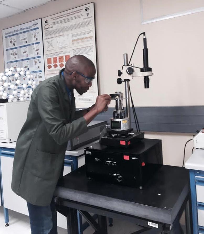
Tshepo Dipheko

PhD student Tshepo Dipheko from South Africa. Credit: RUDN University

What sets chemistry apart from other natural sciences is the ability to get creative and find amazing solutions to long known problems.

A PhD student Tshepo Dipheko from South Africa, instills love for chemistry into people. He doesn’t show it too much, just unwittingly reminds that chemistry surrounds a person absolutely everywhere — it’s in the body, brain, clothing, food and household items. According to the student, it’s impossible to remain indifferent because “Chemistry is everything. We encounter it when drinking coffee or tea, holding a paper cup in our hands, or setting off fireworks on New Year’s Eve”.

Tshepo fell in love with chemistry at school: he was struck not only by the results of colorful chemical reactions, for example, “Pharaoh’s serpent”, but also by the structure of the periodic table and clear chemical equations. Thanks to chemistry, life was ordered by formulas, elements and reactions.

The passion for order and accurate measurements of powders and liquids has moved smoothly to the kitchen. “I’m not the best cook you’ll meet on your way, but I prepare everything with my heart”, says Tshepo. It seems that the student approaches cooking in the same way as preparing the outcome of a reaction in the laboratory: everything is effective, correctly conducted, and the volumes of substances are precisely verified in a scientific way. But he frankly says that “there is no smell of creativity here”. In cooking, you need to respect the principle of all serious scientists in white coats: mix substances following clear instructions without unnecessary amateur activity.

“South Africa doesn’t have enough specialists in chemistry. — Says Tshepo. — Every year we need more and more people with these skills to develop the country’s chemical industry.”

After graduating, Tshepo is waiting for work in the chemical industry and postdoctoral research that opens up the widest opportunities for future scientific activity.

At Last! NASA Successfully Switches to Backup Computer on Hubble Space Telescope
Hubble Space Telescope Inside

Credit: ESA/Hubble (M. Kornmesser)

NASA has successfully switched to backup hardware on the Hubble Space Telescope, including powering on the backup payload computer, on July 15. The switch was performed to compensate for a problem with the original payload computer that occurred on June 13 when the computer halted, suspending science data collection.

The switch included bringing online the backup Power Control Unit (PCU) and the backup Command Unit/Science Data Formatter (CU/SDF) on the other side of the Science Instrument and Command & Data Handling (SI C&DH) unit. The PCU distributes power to the SI C&DH components, and the CU/SDF sends and formats commands and data. In addition, other pieces of hardware onboard Hubble were switched to their alternate interfaces to connect to this backup side of the SI C&DH. Once these steps were completed, the backup payload computer on this same unit was turned on and loaded with flight software and brought up to normal operations mode. 

The Hubble team is now monitoring the hardware to ensure that everything is working properly. The team has also started the process for recovering the science instruments out of their safe mode configuration. This activity is expected to take more than a day as the team runs various procedures and ensures the instruments are at stable temperatures. The team will then conduct some initial calibration of the instruments before resuming normal science operations.

10 reasons why you feel constant hunger

The desire to consume food is the worst enemy of any person who follows a diet. But healthy hunger is a signal that your body needs “fuel” to function properly. And in this case, this fuel is food. If you have missed a meal, then hunger is a completely natural reaction of your body. But if you feel hungry shortly after eating, this is not a good sign. Here are the most common reasons why you constantly feel hungry.

1.Poor sleep

Poor sleep leads to hormonal imbalance. Even if you haven’t slept well one night, it can lead to increased levels of ghrelin (a hormone that stimulates appetite), as well as decreased levels of leptin (a hormone that causes satiety).

2. High levels of stress

When we are under stress, cortisol levels in our body increase. In fact, cortisol makes us crave sugar and high-fat foods.

3. Thyroid problems Excessive hunger is also associated with hyperthyroidism – a common disease affecting the thyroid gland, which means that it is overactive. When thyroid hormone levels are too high, the body’s vital functions are accelerated and you burn energy faster than expected. This explains the increased desire to consume food.

4. Low blood sugar

Low blood sugar or hypoglycaemia means that the glucose in your body has dropped to a very low level. This condition provokes constant hunger. Our brain needs energy to function, and glucose is the fuel it needs. So when your blood sugar goes down, so does your brain activity. Then the brain begins to send signals that the body needs fuel, which is why you begin to feel hungry.

5. Diabetes Type 1 and 2

diabetes can cause frequent bouts of hunger. Under normal circumstances, the body converts sugar from food into a fuel called glucose. But if you have diabetes, the sugar in the food you eat may not get into your tissues. This is the reason you feel constant hunger.

6. Infection with parasites

Sometimes excessive hunger, even shortly after eating, can be a telltale sign of parasite infestation. Worms, especially tapeworms and pinworms, can live in your body for a long time without suspecting their existence.

7. Premenstrual syndrome

Women who are about to menstruate may also notice an increased appetite. Ongoing hormonal changes in the body that occur during the second half of the menstrual cycle and disappear within 1 to 2 days after its onset and can make you feel hungry all the time.

8. Pregnancy

Many expectant mothers experience an increased appetite, which is not bad. In fact, increasing the appetite ensures that the baby gets enough nutrients to grow. It is normal to gain about 4 to 6 pounds in the first trimester, then 1 pound per week in the second and third trimesters.

9. Dehydration

When the body is chronically dehydrated, you may feel constant hunger. In fact, many people even confuse thirst with hunger.

10. Drinking alcohol

A glass of wine or beer with dinner can also be a reason why you constantly feel hungry. Drinking alcohol causes an increase in the hormone ghrelin, which causes a feeling of hunger, even if your stomach is full.

Scientists with a breakthrough to prolong life – we can reach 120 years

Researchers at Bar-Ilan University in Israel have been able to increase the lifespan of mice in the laboratory by 23%. Researchers are now exploring the possibility of prolonging human life, reports The Times of Israel.

The researchers “raise the curtain” and talk about their experiment. They were able to increase the amount of SIRT6 in 250 rodents. It is a protein that affects DNA repair. Thus, the genetic material is reproduced in the body and aging is slowed down.

The head of the laboratory where the study was conducted, Chaim Cohen, said that this was an important scientific discovery. If a way is found to use this method on a person, his life expectancy will increase to 120 years. Now the average life expectancy in the world is 71 years.

In addition, the aging process will slow down. At the same time, the risk of age-related diseases will decrease. One will feel wonderful: one will be in good health and consciousness. Cohen adds that there are many tests ahead so that people can live happily ever after.

Surface of Jupiter’s Moon Europa Churned by Small Impacts – May Currently Have Conditions Suitable for Life
Watery Plumes Jupiter's Moon Europa

This color view of Jupiter’s moon Europa was captured by NASA’s Galileo spacecraft in the late 1990s. Scientists are studying processes that affect the surface as they prepare to explore the icy body. Credit: NASA/JPL-Caltech/SETI Institute

Jupiter’s moon Europa and its global ocean may currently have conditions suitable for life. Scientists are studying processes on the icy surface as they prepare to explore.

It’s easy to see the impact of space debris on our Moon, where the ancient, battered surface is covered with craters and scars. Jupiter’s icy moon Europa withstands a similar trouncing – along with a punch of super-intense radiation. As the uppermost surface of the icy moon churns, material brought to the surface is zapped by high-energy electron radiation accelerated by Jupiter.

NASA-funded scientists are studying the cumulative effects of small impacts on Europa’s surface as they prepare to explore the distant moon with the Europa Clipper mission and study the possibilities for a future lander mission. Europa is of particular scientific interest because its salty ocean, which lies beneath a thick layer of ice, may currently have conditions suitable for existing life. That water may even make its way into the icy crust and onto the moon’s surface.

New research and modeling estimate how far down that surface is disturbed by the process called “impact gardening.” The work, published July 12 in Nature Astronomy, estimates that the surface of Europa has been churned by small impacts to an average depth of about 12 inches (30 centimeters) over tens of millions of years. And any molecules that might qualify as potential biosignatures, which include chemical signs of life, could be affected at that depth.

Europa’s Surface Zoom

In this zoomed-in image of Europa’s surface, captured by NASA’s Galileo mission, the thin, bright layer, visible atop a cliff in the center shows the kind of areas churned by impact gardening. Credit: NASA/JPL-Caltech

That’s because the impacts would churn some material to the surface, where radiation would likely break the bonds of any potential large, delicate molecules generated by biology. Meanwhile, some material on the surface would be pushed downward, where it could mix with the subsurface.

“If we hope to find pristine, chemical biosignatures, we will have to look below the zone where impacts have been gardening,” said lead author Emily Costello, a planetary research scientist at the University of Hawaii at Manoa. “Chemical biosignatures in areas shallower than that zone may have been exposed to destructive radiation.”

Going Deeper

While impact gardening has long been understood to be likely taking place on Europa and other airless bodies in the solar system, the new modeling provides the most comprehensive picture yet of the process. In fact, it is the first to take into account secondary impacts caused by debris raining back down onto Europa’s surface after being kicked up by an initial impact. The research makes the case that Europa’s mid- to high-latitudes would be less affected by the double whammy of impact gardening and radiation.

“This work broadens our understanding of the fundamental processes on surfaces across the solar system,” said Cynthia Phillips, a Europa scientist at NASA’s Jet Propulsion Laboratory in Southern California and a co-author of the study. “If we want to understand the physical characteristics and how planets in general evolve, we need to understand the role impact gardening has in reshaping them.”

Managed by JPL for NASA, Europa Clipper will help develop that understanding. The spacecraft, targeting a 2024 launch, will conduct a series of close flybys of Europa as it orbits Jupiter. It will carry instruments to thoroughly survey the moon, as well as sample the dust and gases that are kicked up above the surface.

More About the Mission

Missions such as Europa Clipper contribute to the field of astrobiology, the interdisciplinary research on the variables and conditions of distant worlds that could harbor life as we know it. While Europa Clipper is not a life-detection mission, it will conduct detailed reconnaissance of Europa and investigate whether the icy moon, with its subsurface ocean, has the capability to support life. Understanding Europa’s habitability will help scientists better understand how life developed on Earth and the potential for finding life beyond our planet.

Managed by Caltech in Pasadena, California, JPL leads the development of the Europa Clipper mission in partnership with APL for NASA’s Science Mission Directorate in Washington. The Planetary Missions Program Office at NASA’s Marshall Space Flight Center in Huntsville, Alabama, executes program management of the Europa Clipper mission.

Spanish company targets mass market with 3D printed “steaks”

The plant-based food industry will continue to grow in double digits in the foreseeable future.

At a time when demand for plant-based meat alternatives is growing, Barcelona-based startup Novameat is using 3D printing technology to produce vegetarian steaks and hopes they will reach the mass market next year.

Novameat plans to sell steaks directly to consumers and companies such as restaurants interested in producing plant-based meat, according to Alexander Campos, the company’s business development manager.

The Spanish company, which developed the technology in 2018, showed how its latest 3D printer produces food at the World Mobile Congress in Barcelona.

“It didn’t taste like a traditional steak, but I was positively surprised because I didn’t expect the texture to be so good,” Ferran Gregory told Reuters after tasting one of the printed steaks at Novameat’s stand at the biggest event. for telecoms in the world.

The company uses 3D technology to test recipes by introducing ingredients through capsules because the process is cheaper than mass production, Campos explains.

When the model is considered successful, it can be produced on a larger scale in larger machines without the use of 3D technology, which can give up to 500 kg per hour. a product that mimics meat, he added.

The goal of the startup is to recreate the muscle fibers in animal meat, but using ingredients that are 100% plant-based. Campos predicts that the plant-based food industry will continue to grow in double digits in the foreseeable future.

The company also announced that it produces the product, which imitates meat, for environmental reasons.

“We are trying to replace animal meat with something that is better for the planet, for ourselves and for the animals,” says Campos.