Africa's Oldest Human Burial Site Uncovered – Child Buried 78,000 Years Ago
Africa’s Oldest Human Burial Site Uncovered – Child Buried 78,000 Years Ago
Panga ya Saidi

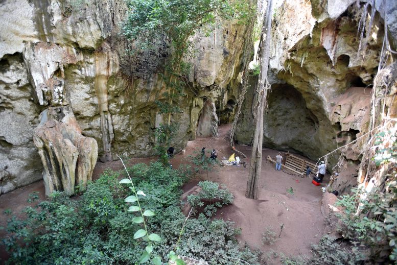
General view of the cave site of Panga ya Saidi. Note trench excavation where burial was unearthed. Credit: Mohammad Javad Shoaee

The discovery of the earliest human burial site yet found in Africa, by an international team including several CNRS researchers,[1] has just been announced in the journal Nature.

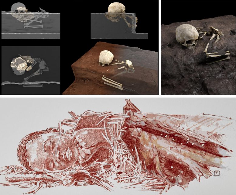
At Panga ya Saidi, in Kenya, north of Mombasa, the body of a three-year-old, dubbed Mtoto (Swahili for ‘child’) by the researchers, was deposited and buried in an excavated pit approximately 78,000 years ago. Through analysis of sediments and the arrangement of the bones, the research team showed that the body had been protected by being wrapped in a shroud made of perishable material, and that the head had likely rested on an object also of perishable material.

Remains and Burial Reconstructions

3D reconstruction of the arrangement of the child’s remains (top), artistic reconstruction of the burial (bottom). Credit: Jorge González / Elena Santos / F. Fuego / MaxPlanck Institute / CENIEH

Though there are no signs of offerings or ochre, both common at more recent burial sites, the funerary treatment given Mtoto suggests a complex ritual that likely required the active participation of many members of the child’s community.
Though Mtoto was a Homo sapiens, the child’s dental morphology, in contrast with that observed in human remains of the same period, preserves certain archaic traits connecting it to distant African ancestors. This apparently confirms that, as has often been posited in recent years, our species has extremely old and regionally diverse roots in the African continent where it arose.

Notes

  1. Participating CNRS researchers hail from the PACEA (CNRS / University of Bordeaux / French Ministry of Culture) and IRAMAT (CNRS / Université Bourgogne Franche-Comté / University of Orléans / Bordeaux Montaigne University) research units. In France, this research was funded by the LaScArBx Laboratory of Excellence (LabEx).

For more on this research, read Africa’s Oldest Human Burial Site Uncovered: 78,000-Year-Old Remains Reveal How Stone Age Populations Interacted With the Dead.

Reference: “Earliest known human burial in Africa” by María Martinón-Torres, Francesco d’Errico, Elena Santos, Ana Álvaro Gallo, Noel Amano, William Archer, Simon J. Armitage, Juan Luis Arsuaga, José María Bermúdez de Castro, James Blinkhorn, Alison Crowther, Katerina Douka, Stéphan Dubernet, Patrick Faulkner, Pilar Fernández-Colón, Nikos Kourampas, Jorge González García, David Larreina, François-Xavier Le Bourdonnec, George MacLeod, Laura Martín-Francés, Diyendo Massilani, Julio Mercader, Jennifer M. Miller, Emmanuel Ndiema, Belén Notario, Africa Pitarch Martí, Mary E. Prendergast, Alain Queffelec, Solange Rigaud, Patrick Roberts, Mohammad Javad Shoaee, Ceri Shipton, Ian Simpson, Nicole Boivin and Michael D. Petraglia, 5 May 2021, Nature.
DOI: 10.1038/s41586-021-03457-8

Simple Surgery Prevents Strokes in Heart Patients – Safe, Inexpensive
Richard Whitlock

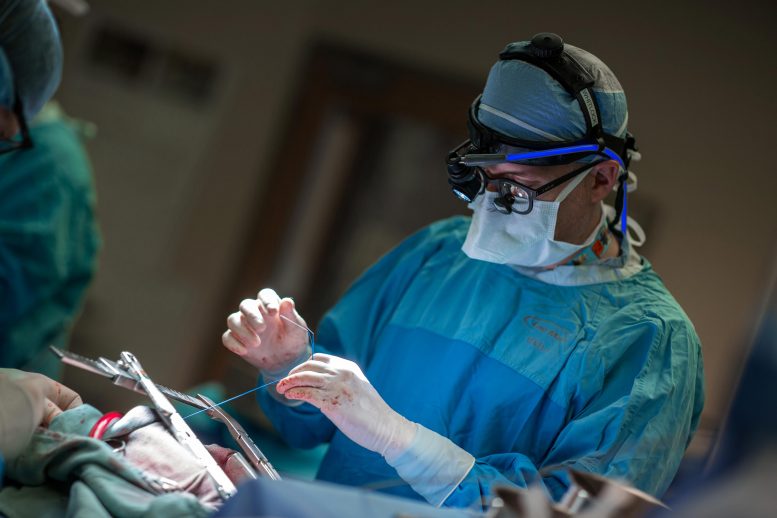
Richard Whitlock, professor of surgery at McMaster University, performing heart surgery. Credit: Hamilton Health Sciences

Removing left atrial appendage cuts the risk of strokes by more than one-third in patients with atrial fibrillation.

A simple surgery saves patients with heart arrhythmia from often-lethal strokes, says a large international study led by McMaster University.

Researchers found that removing the left atrial appendage — an unused, finger-like tissue that can trap blood in the heart chamber and increase the risk of clots — cuts the risk of strokes by more than one-third in patients with atrial fibrillation.

Even better, the reduced clotting risk comes on top of any other benefits conferred by blood-thinner medications patients with this condition are usually prescribed.

“If you have atrial fibrillation and are undergoing heart surgery, the surgeon should be removing your left atrial appendage, because it is a set-up for forming clots. Our trial has shown this to be both safe and effective for stroke prevention,” said Richard Whitlock, first author of the study.

“This is going to have a positive impact on tens of thousands of patients globally.”

Intact Left Atrial Appendage

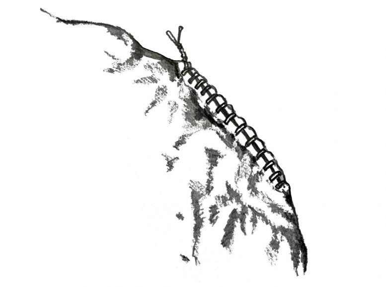
The left atrial appendage, intact. Credit: Heather Borsellino

Whitlock is a scientist at the Population Health Research Institute (PHRI), a joint institute of McMaster University and Hamilton Health Sciences (HHS); a professor of surgery at McMaster, the Canada Research Chair in cardiovascular surgical trials, a cardiac surgeon for HHS, and is supported by a Heart and Stroke Foundation career award.

The co-principal investigator of the study is Stuart Connolly who has also advanced this field by establishing the efficacy and safety of newer blood thinners. He is a professor emeritus of medicine at McMaster, a PHRI senior scientist and an HHS cardiologist.

Left Atrial Appendage After Occlusion

The left atrial appendage after occlusion. Credit: Heather Borsellino

“The results of this study will change practice right away because this procedure is simple, quick and safe for the 15 percent of heart surgery patients who have atrial fibrillation. This will prevent a great burden of suffering due to stroke,” Connolly said.

The study results were fast tracked into publication by The New England Journal of Medicine and presented at the American College of Cardiology conference today.

The study tracked 4,811 people in 27 countries who are living with atrial fibrillation and taking blood thinners. Consenting patients undertaking cardiopulmonary bypass surgery were randomly selected for the additional left atrial appendage occlusion surgery; their outcomes compared with those who only took medicine. They were all followed for a median of four years.

Stuart Connolly

Stuart Connolly. Credit: Population Health Research Institute

Whitlock said it was suspected since the 1940s that blood clots can form in the left atrial appendage in patients with atrial fibrillation, and it made sense to cut this useless structure off if the heart was exposed for other surgery. This is now proven to be true.

Atrial fibrillation is common in elderly people and is responsible for about 25 percent of ischemic strokes which are caused when blood clots block arteries supplying parts of the brain. The average age of patients in the study was 71.

“In the past all we had was medicine. Now we can treat atrial fibrillation with both medicines and surgery to ensure a much better outcome,” said Whitlock.

He said that the current study tested the procedure during cardiac surgery being undertaken for other reasons, but the procedure can also be done through less invasive methods for patients not having heart surgery. He added that future studies to examine that approach will be important.

Whitlock said the left atrial appendage is a leftover from how a person’s heart forms as an embryo and it has little function later in life.

“This is an inexpensive procedure that is safe, without any long-term adverse effects, and the impact is long-term.”

Reference: 15 May 2021, New England Journal of Medicine.

External funding for the study came from the Canadian Institutes of Health Research and the Heart and Stroke Foundation of Canada.

First Images of the Cosmic Web Reveal Unsuspected Presence of Billions of Dwarf Galaxies
Cosmological Simulation of Distant Universe

Figure 1: cosmological simulation of the distant Universe. The image shows the light emitted by hydrogen atoms in the cosmic web in a region roughly 15 million light years across. In addition to the very weak emission from intergalactic gas, a number of point sources can be seen: these are galaxies in the process of forming their first stars. Credit: Jeremy Blaizot / projet SPHINX

  • In the Universe, galaxies are distributed along extremely tenuous filaments of gas millions of light years long separated by voids, forming the cosmic web.
  • The MUSE instrument on the Very Large Telescope has captured an image of several filaments in the early Universe…
  • … revealing the unexpected presence of billions of dwarf galaxies in the filaments

Although the filaments of gas in which galaxies are born have long been predicted by cosmological models, we have so far had no real images of such objects. Now for the first time, several filaments of the ‘cosmic web’ have been directly observed using the MUSE[1] instrument installed on ESO’s Very Large Telescope in Chile. These observations of the early Universe, 1 to 2 billion years after the Big Bang, point to the existence of a multitude of hitherto unsuspected dwarf galaxies. Carried out by an international collaboration led by the Centre de Recherche Astrophysique de Lyon (CNRS/Université Lyon 1/ENS de Lyon), also involving the Lagrange laboratory (CNRS/Université Côte d’Azur/Observatoire de la Côte d’Azur),[2] the study is published in the journal Astronomy & Astrophysics.

2250 Galaxies in Cone of Universe Observed by MUSE

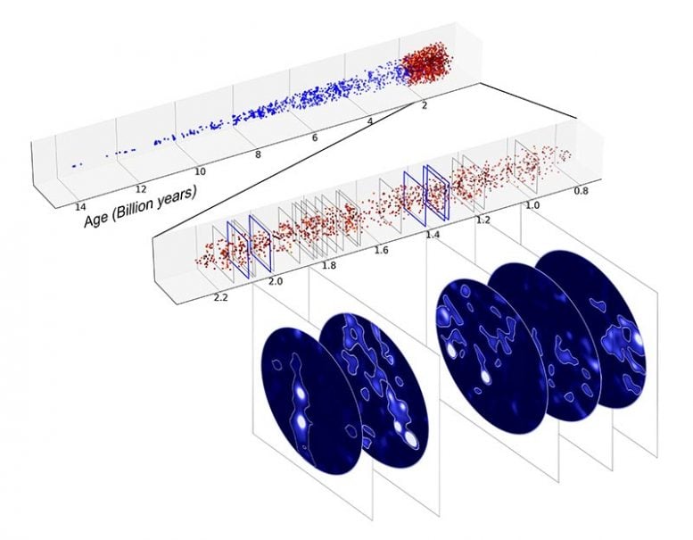
Figure 2: the 2250 galaxies in the ‘cone’ of the Universe observed by MUSE are shown here according to the age of the Universe (in billions of years). The period of the early Universe (0.8 to 2.2 billion years after the Big Bang) explored in this study is shown in red. The 22 regions with galaxy over-density are indicated by grey rectangles. The 5 regions where filaments have been identified most prominently are shown in blue. Credit: Roland Bacon / David Mary

The filamentary structure of hydrogen gas in which galaxies form, known as the cosmic web, is one of the major predictions of the model of the Big Bang and of galaxy formation [figure 1]. Until now, all that was known about the web was limited to a few specific regions, particularly in the direction of quasars, whose powerful radiation acts like car headlights, revealing gas clouds along the line of sight. However, these regions are poorly representative of the whole network of filaments where most galaxies, including our own, were born.  Direct observation of the faint light emitted by the gas making up the filaments was a holy grail which has now been attained by an international team headed by Roland Bacon, CNRS researcher at the Centre de Recherche Astrophysique de Lyon (CNRS/Université Lyon 1/ENS de Lyon).

Hydrogen Filaments Discovered by MUSE

Figure 3: one of the hydrogen filaments (in blue) discovered by MUSE in the Hubble Ultra-Deep Field. It is located in the constellation Fornax at a distance of 11.5 billion light years, and stretches across 15 million light years. The image in the background is from Hubble. Credit: Roland Bacon, David Mary, ESO and NASA

The team took the bold step of pointing ESO’s Very Large Telescope, equipped with the MUSE instrument coupled to the telescope’s adaptive optics system, at a single region of the sky for over 140 hours. Together, the two instruments form one of the most powerful systems in the world.[3] The region selected forms part of the Hubble Ultra-Deep Field, which was until now the deepest image of the cosmos ever obtained. However, Hubble has now been surpassed, since 40% of the galaxies discovered by MUSE have no counterpart in the Hubble images.

Cosmological Simulation of Filament

Figure 4: cosmological simulation of a filament made up of hundreds of thousands of small galaxies. The image on the left shows the emissions produced by all the galaxies as it might be observed in situ. The image on the right shows the filament as it would be seen by MUSE. Even with a very long exposure time, the vast majority of the galaxies cannot be detected individually. However, the light from all these small galaxies is detected as a diffuse background, rather like the Milky Way when seen with the naked eye. Credit: Thibault Garel and Roland Bacon

After meticulous planning, it took eight months to carry out this exceptional observing campaign. This was followed by a year of data processing and analysis, which for the first time revealed light from the hydrogen filaments, as well as images of several filaments as they were one to two billion years after the Big Bang, a key period for understanding how galaxies formed from the gas in the cosmic web [figures 2 et 3]. However, the biggest surprise for the team was when simulations showed that the light from the gas came from a hitherto invisible population of billions of dwarf galaxies spawning a host of stars [figure 4].[4] Although these galaxies are too faint to be detected individually with current instruments, their existence will have major consequences for galaxy formation models, with implications that scientists are only just beginning to explore.

Notes

  1. MUSE, which stands for Multi Unit Spectroscopic Explorer, is a 3D spectrograph designed to explore the distant Universe. The construction of the instrument was led by the Centre de Recherche Astrophysique de Lyon (CNRS/Université Claude Bernard-Lyon 1/ENS de Lyon).
  2. Other French laboratories involved: Laboratoire d’Astrophysique de Marseille (CNRS/Aix-Marseille Université/CNES), Institut de Recherche en Astrophysique et Planétologie (CNRS/Université Toulouse III – Paul Sabatier/CNES).
  3. See ESO press release.
  4. Until now, theory predicted that the light came from the diffuse cosmic ultraviolet background radiation (very weak background radiation produced by all the galaxies and stars) which, by heating the gas in the filaments, causes them to glow.

Reference: “The MUSE Extremely Deep Field: The cosmic web in emission at high redshift” by R. Bacon, D. Mary, T. Garel, J. Blaizot, M. Maseda, J. Schaye, L. Wisotzki, S. Conseil, J. Brinchmann, F. Leclercq, V. Abril-Melgarejo, L. Boogaard, N. F. Bouché, T. Contini, A. Feltre, B. Guiderdoni, C. Herenz, W. Kollatschny, H. Kusakabe, J. Matthee, L. Michel-Dansac, T. Nanayakkara, J. Richard, M. Roth, K. B. Schmidt, M. Steinmetz, L. Tresse, T. Urrutia, A. Verhamme, P. M. Weilbacher, J. Zabl and S. L. Zoutendijk, 18 March 2021, Astronomy & Astrophysic.
DOI: 10.1051/0004-6361/202039887

Scientists Have Figured Out What Triggers Large-Scale Volcanic Eruptions

A lava flow from Hawaii’s Kilauea Volcano enters the ocean near Isaac Hale Beach Park on August 5, 2018. The volcano’s 2018 eruption was its largest in over 200 years. Credit: USGS

Caldera Collapse Increases the Size and Duration of Volcanic Eruptions

Scientists have figured out what triggers large-scale volcanic eruptions and what conditions likely lead to them.

Hawaii’s Kilauea is one of the most active volcanoes in the world. Because of this and its relative ease of accessibility, it is also among the most heavily outfitted with monitoring equipment – instruments that measure and record everything from earthquakes and ground movement to lava volume and advancement.

Kilauea’s 2018 eruption, however, was especially massive. In fact, it was the volcano’s largest eruption in over 200 years. Scientists at NASA’s Jet Propulsion Laboratory in Southern California used the abundance of data collected from this rare event to shed light on the cause of large-scale eruptions like this one and, perhaps more importantly, what mechanisms trigger them.

This image shows the lava flow from the 2018 eruption of Kilauea prior to the collapse of the caldera. Credit: NASA/JPL-Caltech

“Ultimately, what caused this eruption to be so much larger than normal was the collapse of the volcano’s caldera – the large, craterlike depression at the volcano’s summit,” said JPL’s Alberto Roman, lead author of the new study published recently in Nature. “During a caldera collapse, a massive block of rock near the top of the volcano slides down into the volcano. As it slides, gets stuck on the jagged walls around it, and slides some more, the block of rock squeezes out more magma than would ordinarily be expelled.”

This image shows the much larger lava flow that followed the collapse of the caldera. Credit: NASA/JPL-Caltech

But what the science team really wanted to know was what caused the caldera to collapse in the first place – and they found their answer.

The likely culprit? Vents – openings through which lava flows – located a distance away from, and at a much lower elevation than, the volcano’s summit.

“Sometimes, volcanoes erupt at the summit, but an eruption can also occur when lava breaks through vents much lower down the volcano,” said JPL’s Paul Lundgren, co-author of the study. “Eruption through these low-elevation vents likely led to the collapse of the caldera.”

During an eruption, the surface of a volcano deforms, or changes shape. The color bands in the lower-right animation box show those changes from before to midway through Kilauea’s 2018 eruption. The closer the color bands are to one another, the more severe the deformation in that area – much like the contour lines on a topographic map denote rapidly changing altitude. Credit: Credit: NASA/JPL-Caltech

Lundgren compares this type of vent to the spigot on a collapsible water jug you’d take on a camping trip. As the water level drops toward the location of the spigot, the flow of water slows or stops. Likewise, the lower down the volcano a vent (or “spigot”) is located, the longer lava is likely to flow before reaching a stopping point.

A large quantity of magma can be expelled quickly from the chamber (or chambers) beneath the volcano through these vents, leaving the rocky floor and walls of the caldera above the chamber without sufficient support. The rock from the caldera can then collapse into the magma chamber.

As the rock falls, it pressurizes the magma chambers – for Kilauea, the research team identified two of them – increasing the magma flow to the distant vents as well as the total volume of the eruption. The pressurization is akin to squeezing the water jug to force out the last little bit of water.

After developing their model of these eruption processes, taking advantage of the myriad data available from Kilauea, they also compared the model’s predictions to observations from similar eruptions driven by caldera collapse at other volcanoes. The results were consistent. Even though the model doesn’t predict when a volcano is going to erupt, it can provide crucial insight into the likely severity of an eruption once it begins.

“If we see an eruption at a low-elevation vent, that is a red flag or warning that caldera collapse is possible,” said Roman. “Similarly, if we detect earthquakes consistent with the slipping of the caldera rock block, we now know that the eruption will likely be much larger than usual.”

Reference: “Dynamics of large effusive eruptions driven by caldera collapse” by Alberto Roman and Paul Lundgren, 14 April 2021, Nature.
DOI: 10.1038/s41586-021-03414-5

Alien Species Predicted to Increase by 36% Worldwide by 2050

Egyptian goose (Alopochen aegyptiaca) originally from Africa and now established in Central and Western Europe. Credit: Professor Tim Blackburn, UCL

The number of alien (non-native) species, particularly insects, arthropods and birds, is expected to increase globally by 36% by the middle of this century, compared to 2005, finds new research by an international team involving UCL.

Published in Global Change Biology, the study also predicts the arrival of around 2,500 new alien species in Europe, which translates to an increase of 64% for the continent over the 45-year period.

The research team led by the German Senckenberg Biodiversity and Climate Research Centre hope it should be possible to reduce this number with stricter biosecurity regulations.

Alien species are those that humans have moved around the world to places where they do not naturally occur. More than 35,000 such species had been recorded by 2005 (the date of the most recent comprehensive global catalogue). Some of these aliens can go on to become invasive, with damaging impacts to ecosystems and economies. Alien species are one of the main drivers of extinctions of animals and plants.

Co-author Professor Tim Blackburn (UCL Centre for Biodiversity & Environment Research and the Institute of Zoology, ZSL) said: “Our study predicts that alien species will continue to be added to ecosystems at high rates through the next few decades, which is concerning as this could contribute to harmful biodiversity change and extinction.

“But we are not helpless bystanders: with a concerted global effort to combat this, it should be possible to slow down or reverse this trend.”

Box tree moth, native to east Asia and now found across Europe. Credit: Professor Tim Blackburn, UCL

For the study, the research team developed a mathematical model to calculate for the first time how many more aliens would be expected by 2050, based on estimated sizes of source pools (the species that could end up becoming invasive) and dynamics of historical invasions, under a ‘business-as-usual’ scenario that assumes a continuation of current trends.

The model predicts a 36% increase in the number of alien plant and animal species worldwide by 2050, compared to 2005 levels.

The study identifies high levels of variation between regions. The largest increase is expected in Europe, where the number of alien species will increase by 64% by the middle of the century. Additional alien hotspots are predicted to include temperate latitudes of Asia, North America, and South America. The lowest relative increase in alien species is expected in Australia.

Europe will also see the largest increase in absolute numbers of alien species, with around 2,500 new aliens predicted.

Lead author Dr Hanno Seebens (Senckenberg Biodiversity and Climate Research Centre, Germany) said: “These will primarily include rather inconspicuous new arrivals such as insects, molluscs, and crustaceans. In contrast, there will be very few new alien mammal species such as the well-known raccoon.”

Co-author Dr Franz Essl (University of Vienna) added: “Increases are expected to be particularly large for insects and other arthropods, such as arachnids and crustaceans. We predict the number of aliens from these groups to increase in every region of the world by the middle of the century – by almost 120% in the temperate latitudes of Asia.”

The study also predicts that the rate of arrival of alien species will continue to increase, at least in some animal groups. Globally, by 2050, alien arthropod and bird species in particular will arrive faster than before, compared to the period 1960 – 2005. In Europe, the rate of new alien arrivals is expected to increase for all plant and animal groups except mammals.

Neither a reversal nor even a slowdown in the spread of alien species is in sight, as global trade and transport are expected to increase in the coming decades, allowing many species to infiltrate new habitats as stowaways.

Dr Seebens said: “We will not be able to entirely prevent the introduction of alien species, as this would mean severe restrictions in international trade.

“However, stricter regulations and their rigorous enforcement could greatly slow the flow of new species. The benefits of such measures have been shown in some parts of the world. Regulations are still comparatively lax in Europe, and so there is great potential here for new measures to curtail the arrival of new aliens.”

Reference: “Projecting the continental accumulation of alien species through to 2050” by Hanno Seebens, Sven Bacher, Tim M. Blackburn, César Capinha, Wayne Dawson, Stefan Dullinger, Piero Genovesi, Philip E. Hulme, Mark van Kleunen, Ingolf Kühn, Jonathan M. Jeschke, Bernd Lenzner, Andrew M. Liebhold, Zarah Pattison, Jan Pergl, Petr Pyšek, Marten Winter and Franz Essl, 1 October 2020, Global Change Biology.
DOI: 10.1111/gcb.15333

Carb-Eating Bacteria Under Viral Threat: Scientists Discover New Group of Viruses That Attack Bacteria in Our Guts

Assorted Fresh Bread

Scientists characterize previously unknown gut reactions.

Strictly speaking, humans cannot digest complex carbohydrates — that’s the job of bacteria in our large intestines. UC Riverside scientists have just discovered a new group of viruses that attack these bacteria.

The viruses, and the way they evade counterattack by their bacterial hosts, are described in a paper published in Cell Reports.

Bacterioides can constitute up to 60% of all the bacteria living in a human’s large intestine, and they’re an important way that people get energy. Without them, we’d have a hard time digesting bread, beans, vegetables, or other favorite foods. Given their significance, it is surprising that scientists know so little about viruses that prey on Bacteroides

“This is largely unexplored territory,” said microbiologist Patrick Degnan, an assistant professor of microbiology and plant pathology, who led the research. 

To find a virus that attacks Bacteroides, Degnan and his team analyzed a collection of bacterial genomes, where viruses can hide for numerous generations until something triggers them to replicate, attack, and leave their host. This viral lifestyle is not without risk as over time mutations could occur that prevent the virus from escaping its host.

On analyzing the genome of Bacteroides vulgatus, Degnan’s team found DNA belonging to a virus they named BV01. However, determining whether the virus is capable of escaping, or re-infecting its host, proved challenging.

Bacteriophage Reconstructed Microscopy Image

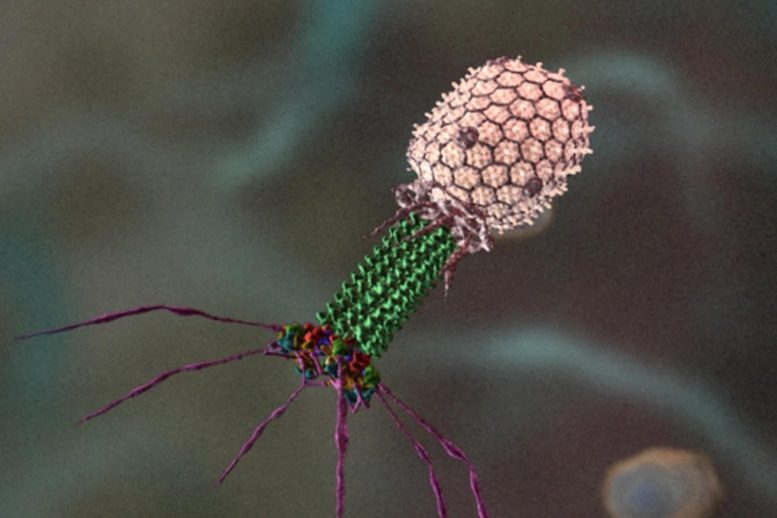
Reconstructed microscopy image of a bacteriophage, which is a virus that attacks bacteria. Credit: Purdue University and Seyet LLC

“We tried every trick we could think of. Nothing in the laboratory worked until we worked with a germ-free mouse model,” Degnan said. “Then, the virus jumped.” 

This was possible due to Degnan’s collaboration with UCR colleague, co-author and fellow microbiologist Ansel Hsiao.

This result suggests conditions in mammalian guts act as a trigger for BV01 activity. The finding underscores the importance of both in vitro and in vivo experiments for understanding the biology of microbes. 

Looking for more information about the indirect effect of this bacterial virus might have on humans, Degnan’s team determined that when BV01 infects a host cell, it disrupts how that cell normally behaves. 

“Over 100 genes change how they get expressed after infection,” Degnan said. 

Two of the altered genes that stood out to the researchers are both responsible for deactivating bile acids, which are toxic to microbes. The authors speculate that while this possibly alters the sensitivity of the bacteria to bile acids, it also may influence the ability of the bacteria to be infected by other viruses.

“This virus can go in and change the metabolism of these bacteria in human guts that are so key for our own metabolism,” Degnan said. 

Though the full extent of BV01 infection is not yet known, scientists believe viruses that change the abundance and activity of gut bacteria contribute to human health and disease. One area for future studies will involve the effect of diet on BV01 and viruses like it, as certain foods can cause our bodies to release more bile.

Degnan also notes that BV01 is only one of a group of viruses his team identified that function in similar ways. The group, Salyersviridae, is named after famed microbiologist Abigail Salyers whose work on intestinal bacteria furthered the science of antibiotic resistance.

Further research is planned to understand the biology of these viruses. 

“It’s been sitting in plain sight, but no one has characterized this important group of viruses that affect what’s in our guts until now,” Degnan said.

Reference: “Infection with Bacteroides Phage BV01 Alters the Host Transcriptome and Bile Acid Metabolism in a Common Human Gut Microbe” by Danielle E. Campbell, Lindsey K. Ly and Jason M. R, 15 September 2020, Cell Reports.
DOI: 10.1016/j.celrep.2020.108142

No Joke: Pigs and Rodents Can Breathe Through Their Butts

Happy Pig on Floor

Rodents and pigs share with certain aquatic organisms the ability to use their intestines for respiration, finds a study publishing May 14th in the journal Med. The researchers demonstrated that the delivery of oxygen gas or oxygenated liquid through the rectum provided vital rescue to two mammalian models of respiratory failure.

“Artificial respiratory support plays a vital role in the clinical management of respiratory failure due to severe illnesses such as pneumonia or acute respiratory distress syndrome,” says senior study author Takanori Takebe (@TakebeLab) of the Tokyo Medical and Dental University and the Cincinnati Children’s Hospital Medical Center. “Although the side effects and safety need to be thoroughly evaluated in humans, our approach may offer a new paradigm to support critically ill patients with respiratory failure.”

Several aquatic organisms have evolved unique intestinal breathing mechanisms to survive under low-oxygen conditions using organs other than lungs or gills. For example, sea cucumbers, freshwater fish called loaches, and certain freshwater catfish use their intestines for respiration. But it has been heavily debated whether mammals have similar capabilities.

In the new study, Takebe and his collaborators provide evidence for intestinal breathing in rats, mice, and pigs. First, they designed an intestinal gas ventilation system to administer pure oxygen through the rectum of mice. They showed that without the system, no mice survived 11 minutes of extremely low-oxygen conditions. With intestinal gas ventilation, more oxygen reached the heart, and 75% of mice survived 50 minutes of normally lethal low-oxygen conditions.

Because the intestinal gas ventilation system requires abrasion of the intestinal muscosa, it is unlikely to be clinically feasible, especially in severely ill patients–so the researchers also developed a liquid-based alternative using oxygenated perfluorochemicals. These chemicals have already been shown clinically to be biocompatible and safe in humans.

The intestinal liquid ventilation system provided therapeutic benefits to rodents and pigs exposed to non-lethal low-oxygen conditions. Mice receiving intestinal ventilation could walk farther in a 10% oxygen chamber, and more oxygen reached their heart, compared to mice that did not receive intestinal ventilation. Similar results were evident in pigs. Intestinal liquid ventilation reversed skin pallor and coldness and increased their levels of oxygen, without producing obvious side effects. Taken together, the results show that this strategy is effective in providing oxygen that reaches circulation and alleviates respiratory failure symptoms in two mammalian model systems.

With support from the Japan Agency for Medical Research and Development to combat the coronavirus disease 2019 (COVID-19) pandemic, the researchers plan to expand their preclinical studies and pursue regulatory steps to accelerate the path to clinical translation.

“The recent SARS-CoV-2 pandemic is overwhelming the clinical need for ventilators and artificial lungs, resulting in a critical shortage of available devices, and endangering patients’ lives worldwide,” Takebe says. “The level of arterial oxygenation provided by our ventilation system, if scaled for human application, is likely sufficient to treat patients with severe respiratory failure, potentially providing life-saving oxygenation.”

Reference: “Mammalian enteral ventilation ameliorates respiratory failure” by Ryo Okabe, Toyofumi F. Chen-Yoshikawa, Yosuke Yoneyama, Yuhei Yokoyama, Satona Tanaka, Akihiko Yoshizawa, Wendy L. Thompson, Gokul Kannan, Eiji Kobayashi, Hiroshi Date and Takanori Takebe, 14 May 2021, Med.
DOI: 10.1016/j.medj.2021.04.004

This work was supported by Research Program on Emerging and Re-emerging Infectious Diseases, Research Projects on COVID-19, from the Japan Agency for Medical Research and Development, and AMED The Translational Research program and AMED Program for technological innovation of regenerative medicine.

X-ray Experiments and Machine Learning Innovation Could Trim Years off Battery R&D
Battery Informatics Lab 1070

Staff engineer Bruis van Vlijmen is seen working inside the Battery Informatics Lab 1070 in the Arrillaga Science Center, Bldg. 57. Credit: Jacqueline Orrell/SLAC National Accelerator Laboratory

An X-ray instrument at Berkeley Lab contributed to a battery study that used an innovative approach to machine learning to speed up the learning curve about a process that shortens the life of fast-charging lithium batteries.

Researchers used Berkeley Lab’s Advanced Light Source, a synchrotron that produces light ranging from the infrared to X-rays for dozens of simultaneous experiments, to perform a chemical imaging technique known as scanning transmission X-ray microscopy, or STXM, at a state-of-the-art ALS beamline dubbed COSMIC

Researchers also employed “in situ” X-ray diffraction at another synchrotron – SLAC’s Stanford Synchrotron Radiation Lightsource – which attempted to recreate the conditions present in a battery, and additionally provided a many-particle battery model. All three forms of data were combined in a format to help the machine-learning algorithms learn the physics at work in the battery.

While typical machine-learning algorithms seek out images that either do or don’t match a training set of images, in this study the researchers applied a deeper set of data from experiments and other sources to enable more refined results. It represents the first time this brand of “scientific machine learning” was applied to battery cycling, researchers noted. The study was published recently in Nature Materials.

The study benefited from an ability at the COSMIC beamline to single out the chemical states of about 100 individual particles, which was enabled by COSMIC’s high-speed, high-resolution imaging capabilities. Young-Sang Yu, a research scientist at the ALS who participated in the study, noted that each selected particle was imaged at about 50 different energy steps during the cycling process, for a total of 5,000 images. 

The data from ALS experiments and other experiments were combined with data from fast-charging mathematical models, and with information about the chemistry and physics of fast charging, and then incorporated into the machine-learning algorithms.

“Rather than having the computer directly figure out the model by simply feeding it data, as we did in the two previous studies, we taught the computer how to choose or learn the right equations, and thus the right physics,” said Stanford postdoctoral researcher Stephen Dongmin Kang, a study co-author.

Patrick Herring, senior research scientist for Toyota Research Institute, which supported the work through its Accelerated Materials Design and Discovery program, said, “By understanding the fundamental reactions that occur within the battery, we can extend its life, enable faster charging, and ultimately design better battery materials.”

Reference: “Fictitious phase separation in Li layered oxides driven by electro-autocatalysis” by Jungjin Park, Hongbo Zhao, Stephen Dongmin Kang, Kipil Lim, Chia-Chin Chen, Young-Sang Yu, Richard D. Braatz, David A. Shapiro, Jihyun Hong, Michael F. Toney, Martin Z. Bazant and William C. Chueh, 8 March 2021, Nature Materials.
DOI: 10.1038/s41563-021-00936-1

Deep Space Listening: 6000 Days of Research to Hear Continuous Gravitational Waves
Rapidly Rotating Neutron Stars

Rapidly rotating neutron stars may be “humming” continuous gravitational waves. Credit: K. Wette

Remember the days before working from home? It’s Monday morning, you’re running late to beat the traffic, and you can’t find your car keys. What do you do? You might try moving from room to room, casting your eye over every flat surface, in the hope of spotting the missing keys. Of course, this assumes that they are somewhere in plain sight; if they’re hidden under a newspaper, or fallen behind the sofa, you’ll never spot them. Or you might be so convinced that you last saw the keys in the kitchen and search for them there: inside every cupboard, the microwave, dishwasher, back of the fridge, etc. Of course, if you left them on your bedside table, upending the kitchen is doomed to failure. So, which is the best strategy?
 
Scientists face a similar conundrum in the hunt for gravitational waves—ripples in the fabric of space and time—from rapidly spinning neutron stars. These stars are the densest objects in the Universe and, provided they’re not perfectly spherical, emit a very faint “hum” of continuous gravitational waves. Hearing this “hum” would allow scientists to peer deep inside a neutron star and discover its secrets, yielding new insights into the most extreme states of matter. However, our very sensitive “ears”—4-kilometer-sized detectors using powerful lasers—haven’t heard anything yet.
 
Part of the challenge is that, like the missing keys, scientists aren’t sure of the best search strategy. Most previous studies have taken the “room-to-room” approach, trying to find continuous gravitational waves in as many different places as possible. But this means you can only spend a limited amount of time listening for the tell-tale “hum” in any one location—in the same way that you can only spend so long staring at your coffee table, trying to discern a key-shaped object. And since the “hum” is very quiet, there’s a good chance you won’t even hear it.
 
In a study recently published in Physical Review D, a team of scientists, led by postdoctoral researcher Karl Wette from the ARC Centre of Excellence for Gravitational Wave Discovery (OzGrav) at the Australian National University, tried the “where else could they be but the kitchen?” approach.
 
Wette explains: “We took an educated guess at a specific location where continuous gravitational waves might be, based in part on what we already know about pulsars—they’re like neutron stars but send out radio waves instead of continuous gravitational waves. We hypothesized that there would be continuous gravitational waves detected near pulsar radio waves.” Just like guessing that your missing keys will probably be close to your handbag or wallet.
 
Using existing observational data, the team spent a lot of time searching in this location (nearly 6000 days of computer time!) listening carefully for that faint “hum.” They also used graphic processing units—specialist electronics normally used for computer games—making their algorithms run super-fast.

“Our search was significantly more sensitive than any previous search for this location,” says Wette. “Unfortunately, we didn’t hear anything, so our guess was wrong this time. It’s back to the drawing board for now, but we’ll keep listening.”

Reference: “Deep exploration for continuous gravitational waves at 171–172 Hz in LIGO second observing run data” by Karl Wette, Liam Dunn, Patrick Clearwater and Andrew Melatos, 23 March 2021, Physical Review D.
DOI: 10.1103/PhysRevD.103.083020

The Surprising
The Surprising “Secret” Lens Making Method Used by the “Father of Microbiology” Discovered After 300 Years
Leeuwenhoek Microscope

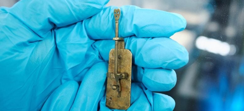
A Van Leeuwenhoek microscope. Credit: Utrecht University/Rijksmuseum Boerhaave/TU Delft

A microscope used by Antoni van Leeuwenhoek to conduct pioneering research contains a surprisingly ordinary lens, as new research by Rijksmuseum Boerhaave Leiden and TU Delft shows. It is a remarkable finding, because Van Leeuwenhoek (1632-1723) led other scientists to believe that his instruments were exceptional. Consequently, there has been speculation about his method for making lenses for more than three centuries. The results of this study were published in Science Advances today (May 14, 2021).

Previous research carried out in 2018 already indicated that some of Van Leeuwenhoek’s microscopes contained common ground lenses. Researchers have now examined a particularly highly magnifying specimen, from the collection of the University Museum Utrecht. Although it did contain a different type of lens, the great surprise was that the lens-making method used was a common one.

Reactor Institute Delft

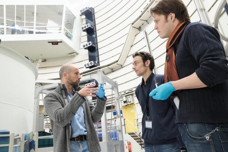
TU Delft researcher Lambert van Eijck and curators Tiemen Cocquyt and Auke Gerrits of Rijksmuseum Boerhaave at the Reactor Institute Delft in The Netherlands. Credit: TU Delft

Pioneering but secretive

With his microscopes, Antoni van Leeuwenhoek saw a whole new world full of minute life which nobody had ever suspected could exist. He was the first to observe unicellular organisms, which is why he is called the father of microbiology. The detail of his observations was unprecedented and was only superseded over a century after his death.

His contemporaries were very curious about the lenses with which Van Leeuwenhoek managed to achieve such astounding feats. Van Leeuwenhoek, however, was very secretive about it, suggesting he had found a new way of making lenses. It now proves to have been an empty boast, at least as far as the Utrecht lens is concerned. This became clear when the researchers from Rijksmuseum Boerhaave Leiden and TU Delft subjected the Utrecht microscope to neutron tomography. It enabled them to examine the lens without opening the valuable microscope and destroy it in the process. The instrument was placed in a neutron beam at the Reactor Institute Delft, yielding a three-dimensional image of the lens.

Hooke Lens

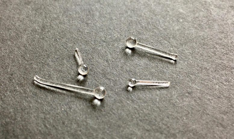
Microscope lenses reconstructed according to the method of Robert Hooke, which Antoni van Leeuwenhoek also used for his highly magnifying microscopes. Credit: Rijksmuseum Boerhaave/TU Delft

Small globule

This lens turned out to be a small globule, and its appearance was consistent with a known production method used in Van Leeuwenhoek’s time. The lens was very probably made by holding a thin glass rod in the fire, so that the end curled up into a small ball, which was then broken off the glass rod.

This method was described in 1678 by another influential microscopist, the Englishman Robert Hooke, which inspired other scientists to do the same. Van Leeuwenhoek, too, may have taken his lead from Hooke. The new discovery is ironical, because it was in fact Hooke who was very curious to learn more about Van Leeuwenhoek’s ‘secret’ method.

The new study shows that Van Leeuwenhoek obtained extraordinary results with strikingly ordinary lens production methods.

Van Leeuwenhoek Microscope

The Van Leeuwenhoek microscope in question, property of the University Museum of Utrecht University. Credit: Utrecht University/Rijksmuseum Boerhaave/TU Delft

Reference: 14 May 2021, Science Advances.

Quantum Leap for Quantum Computing: Ion Beams Create Chains of Closely Coupled Qubits
Ion Beams Qubits Diamond

Ion beams can create chains of closely coupled quantum bits (qubits) based on nitrogen-vacancy “color centers” in diamond for use in quantum computing hardware. The honeycomb pattern in the photo shows the difference between areas exposed to the beam (darker) and masked-off areas. Results indicate it should be possible to create 10,000 coupled qubits over a distance of about the width of a human hair, an unrivaled number and density of qubits. Credit: Susan Brand/Berkeley Lab)

A new way to form self-aligned ‘color centers’ promises scalability to over 10,000 qubits for applications in quantum sensing and quantum computing.

Achieving the immense promise of quantum computing requires new developments at every level, including the computing hardware itself.  A Lawrence Berkeley National Laboratory (Berkeley Lab)-led international team of researchers has discovered a way to use ion beams to create long strings of “color center” qubits in diamond. Their work is detailed in the journal Applied Physics Letters.

The authors includes several from Berkeley Lab: Arun Persaud, who led the study, and Thomas Schenkel, head of the Accelerator Technology and Applied Physics (ATAP) Division’s Fusion Science & Ion Beam Technology Program, as well as Casey Christian (now with Berkeley Lab’s Physics Division), Edward Barnard of Berkeley Lab’s Molecular Foundry, and ATAP affiliate Russell E. Lake.

Creating large numbers of high-quality quantum bits (qubits), in close enough proximity for coupling to each other, is one of the great challenges of quantum computing.  Collaborating with colleagues worldwide, the team has been exploring the use of ion beams to create artificial color centers in diamond for use as qubits.

Color centers are microscopic defects – departures from the rigorous lattice structure of a crystal, such as diamond. The type of defect that is of specific interest for qubits is a nitrogen atom next to a vacancy, or empty space, in a diamond lattice. (Nitrogen is commonly found in the crystal lattice of diamond, which is primarily a crystalline form of carbon, and can contribute to the color of the stone.)

When excited by the rapid energy deposition of a passing ion, nitrogen-vacancy centers can form in the diamond lattice. The electron and nuclear spins of nitrogen-vacancy centers and the adjacent carbon atoms can all function as solid-state qubits, and the crystal lattice can help protect their coherence and mutual entanglement.

Arun Persaud

ATAP Division staff scientist Arun Persaud, principal investigator of this effort. Credit: Marilyn Sargent/Berkeley Lab

The result is a physically durable system that does not have to be used in a cryogenic environment, which are attractive attributes for quantum sensors and also for qubits in this type of solid-state quantum computer. However, making enough qubits, and making them close enough to each other, has been a challenge.

When swift (high-energy) heavy ions such as the beams this team used – gold ions with a kinetic energy of about one billion electron volts – pass through a material, such as nitrogen-doped diamond, they leave a trail of nitrogen-vacancy centers along their tracks. Color centers were found to form directly, without need for further annealing (heat treatment). What’s more, they formed all along the ion tracks, rather than only at the end of the ion range as had been expected from earlier studies with lower-energy ions. In these straight “percolation chains,” color-center qubits are aligned over distances of tens of microns, and are just a few nanometers from their nearest neighbors. A technique developed by Berkeley Lab’s Molecular Foundry measured color centers with depth resolution.

The work on qubit synthesis far from equilibrium was supported by the Department of Energy’s Office of Science. The next step in the research will be to physically cut out a group of these color centers – which are like a series of beads on a string – and show that they are indeed so closely coupled that they can be used as quantum registers.

Results published in the current article show that it will be possible to form quantum registers with up to about 10,000 coupled qubits – two orders of magnitude greater than achieved thus far with the complementary technology of ion-trap qubits – over a distance of about 50 microns (about the width of a human hair).

“Interactions of swift heavy ions with materials have been studied for decades for a variety of purposes, including the behavior of nuclear materials and the effects of cosmic rays on electronics,” said Schenkel.

He added that researchers worldwide have sought to make quantum materials by artificially inducing color centers in diamond. “The solid-state approaches to quantum computing hardware scale beautifully, but integration has been a challenge. This is the first time that direct formation of color-center qubits along strings has been observed.”

The stars, like diamonds

On a miniscule and ephemeral scale (nanometers and picoseconds) the deposition of energy by the ion beams produces a state of high temperature, which Schenkel likens to the surface of the sun, in the 5000 K range, and pressure. Besides knocking carbon atoms out of the crystal lattice of diamond, this effect could enable fundamental studies of exotic states of transient warm dense matter, a state of matter that is present in many stars and large planets and which is difficult to study directly on Earth.

It might also enable formation of novel qubits with tailored properties that cannot be formed with conventional methods. “This opens a new direction for expanding our ability to form quantum registers,” said Schenkel.

iP2 Beamline Target Chamber

Clockwise from bottom left: ATAP Division postdoctoral scholars Sahel Hakimi and Lieselotte Obst-Huebl, and staff scientists Kei Nakamura and Qing Ji, are shown at the target chamber of the iP2 beamline. A high-intensity, short-focal-length beam line, now under construction with DOE Office of Fusion Energy Sciences support, iP2 will be used for laser-based ion acceleration at the Berkeley Lab Laser Accelerator Center (BELLA). Laser-plasma ion acceleration offers the hope of performing many functions using a facility substantially smaller than conventional accelerators. Credit: Thor Swift/Berkeley Lab

Currently, color-center strings are formed with beams from large particle accelerators, such as the one at the German laboratory GSI that was used in this research. In the future, they might be made using compact laser-plasma accelerators like the ones being developed at the Berkeley Lab Laser Accelerator (BELLA) Center. 

The BELLA Center is actively developing its ion-acceleration capabilities with funding by the DOE Office of Science. These capabilities will be used as part of LaserNetUS. Ion pulses from laser-plasma acceleration are very intense and greatly expand our ability to form transient states of highly excited and hot materials for qubit synthesis under novel conditions.

More facets in materials science far from equilibrium

The process of creating these color centers is interesting in its own right and has to be better understood as part of further progress in these applications. The details of how an intense ion beam deposits energy as it traverses the diamond samples, and the exact mechanism by which this leads to color-center formation, hold exciting prospects for further research.

“This work demonstrates both the discovery science opportunities and the potential for societally transformative innovations enabled by the beams from accelerators,” says ATAP Division Director Cameron Geddes. “With accelerators, we create unique states of matter and new capabilities that are not possible by other means.”

Reference: “Direct formation of nitrogen-vacancy centers in nitrogen doped diamond along the trajectories of swift heavy ions” by Russell E. Lake, Arun Persaud, Casey Christian, Edward S. Barnard, Emory M. Chan, Andrew A. Bettiol, Marilena Tomut, Christina Trautmann and Thomas Schenkel, 24 February 2021, Applied Physics Letters.
DOI: 10.1063/5.0036643

The Oldest Centrosaurine: Newly Described Horned Dinosaur From New Mexico Was the Earliest of Its Kind
Menefeeceratops sealeyi

With a frilled head and beaked face, Menefeeceratops sealeyi, discovered in New Mexico, lived 82 million years ago. It predated its better-known relative, Triceratops. Credit: Sergey Krasovskiy

With a frilled head and beaked face, Menefeeceratops sealeyi lived 82 million years ago, predating its relative, Triceratops. Researchers including Peter Dodson, of the School of Veterinary Medicine, and Steven Jasinski, who recently earned his doctorate from the School of Arts & Sciences, describe the find.

A newly described horned dinosaur that lived in New Mexico 82 million years ago is one of the earliest known ceratopsid species, a group known as horned or frilled dinosaurs. Researchers reported their find in a publication in the journal PalZ (Paläontologische Zeitschrift).

Menefeeceratops sealeyi adds important information to scientists’ understanding of the evolution of ceratopsid dinosaurs, which are characterized by horns and frills, along with beaked faces. In particular, the discovery sheds light on the centrosaurine subfamily of horned dinosaurs, of which Menefeeceratops is believed to be the oldest member. Its remains offer a clearer picture of the group’s evolutionary path before it went extinct at the end of the Cretaceous.

Steven Jasinski, who recently completed his Ph.D. in Penn’s Department of Earth and Environmental Science in the School of Arts & Sciences, and Peter Dodson of the School of Veterinary Medicine and Penn Arts & Sciences, collaborated on the work, which was led by Sebastian Dalman of the New Mexico Museum of Natural History and Science. Spencer Lucas and Asher Lichtig of the New Mexico Museum of Natural History and Science in Albuquerque were also part of the research team.

“There has been a striking increase in our knowledge of ceratopsid diversity during the past two decades,” says Dodson, who specializes in the study of horned dinosaurs. “Much of that has resulted from discoveries farther north, from Utah to Alberta. It is particularly exciting that this find so far south is significantly older than any previous ceratopsid discovery. It underscores the importance of the Menefee dinosaur fauna for the understanding of the evolution of Late Cretaceous dinosaur faunas throughout western North America.”

The fossil specimen of the new species, including multiple bones from one individual, was originally discovered in 1996 by Paul Sealey, a research associate of the New Mexico Museum of Natural History and Science, in Cretaceous rocks of the Menefee Formation in northwestern New Mexico. A field crew from the New Mexico Museum of Natural History and Science collected the specimen. Tom Williamson of the New Mexico Museum of Natural History and Science briefly described it the following year, and recent research on other ceratopsid dinosaurs and further preparation of the specimen shed important new light on the fossils.

Based on the latest investigations, researchers determined the fossils represent a new species. The genus name Menefeeceratops refers to the rock formation in which it was discovered, the Menefee Formation, and to the group of which the species is a part, Ceratopsidae. The species name sealeyi honors Sealey, who unearthed the specimen.

Menefeeceratops is related to but predates Triceratops, another ceratopsid dinosaur. However Menefeeceratops was a relatively small member of the group, growing to around 13 to 15 feet long, compared to Triceratops, which could grow to up to 30 feet long.

Horned dinosaurs were generally large, rhinoceros-like herbivores that likely lived in groups or herds. They were significant members of Late Cretaceous ecosystems in North America. “Ceratopsids are better known from various localities in western North America during the Late Cretaceous near the end of the time of dinosaurs,” says Jasinski. “But we have less information about the group, and their fossils are rarer, when you go back before about 79 million years ago.”

Although bones of the entire dinosaur were not recovered, a significant amount of the skeleton was preserved, including parts of the skull and lower jaws, forearm, hindlimbs, pelvis, vertebrae, and ribs. These bones not only show the animal is unique among known dinosaur species but also provide additional clues to its life history. For example, the fossils show evidence of a potential pathology, resulting from a minor injury or disease, on at least one of the vertebrae near the base of its spinal column.

Some of the key features that distinguish Menefeeceratops from other horned dinosaurs involve the bone that make up the sides of the dinosaur’s frill, known as the squamosal. While less ornate than those of some other ceratopsids, Menefeeceratops’ squamosal has a distinct pattern of concave and convex parts.

Comparing features of Menefeeceratops with other known ceratopsid dinosaurs helped the research team trace its evolutionary relationships. Their analysis places Menefeeceratops sealeyi at the base of the evolutionary tree of the centrosaurines subfamily, suggesting that not only is Menefeeceratops one of the oldest known centrosaurine ceratopsids, but also one of the most basal evolutionarily.

Menefeeceratops was part of an ancient ecosystem with numerous other dinosaurs, including the recently recognized nodosaurid ankylosaur Invictarx and the tyrannosaurid Dynamoterror, as well as hadrosaurids and dromaeosaurids.

“Menefeeceratops was part of a thriving Cretaceous ecosystem in the southwestern United States with dinosaurs that predated a lot of the more well-known members closer to end of the Cretaceous,” says Jasinski.

While relatively less work has been done collecting dinosaurs in the Menefee Formation to date, the researchers hope that more field work and collecting in these areas, together with new analyses, will turn up more fossils of Menefeeceratops and ensure a better understanding of the ancient ecosystem of which it was part.

Reference: “The oldest centrosaurine: a new ceratopsid dinosaur (Dinosauria: Ceratopsidae) from the Allison Member of the Menefee Formation (Upper Cretaceous, early Campanian), northwestern New Mexico, USA” by Sebastian G. Dalman, Spencer G. Lucas, Steven E. Jasinski, Asher J. Lichtig and Peter Dodson, 10 May 2021, PalZ.
DOI: 10.1007/s12542-021-00555-w

Peter Dodson is a professor of anatomy in the School of Veterinary Medicine and a professor of earth and environmental science in the School of Arts & Sciences at the University of Pennsylvania.

Steven E. Jasinski is a curator of paleontology and geology at the State Museum of Pennsylvania and corporate faculty at Harrisburg University of Science and Technology. He earned his doctoral degree in the Department of Earth and Environmental Science in the University of Pennsylvania’s School of Arts & Sciences.

Sebastian G. Dalman is a research associate at the New Mexico Museum of Natural History and Science in Albuquerque.

Spencer G. Lucas is a curator of paleontology at the New Mexico Museum of Natural History and Science in Albuquerque.

Asher J. Lichtig is a research associate at the New Mexico Museum of Natural History and Science in Albuquerque.

Jasinski was supported by Geo. L. Harrison and Benjamin Franklin fellowships while attending the University of Pennsylvania. The research was also partially funded by a Walker Endowment Research Grant and a University of Pennsylvania Paleontology Research Grant.

One-Third of Global Food Production Threatened by Climate Change
Global Food Production Emissions Comparison

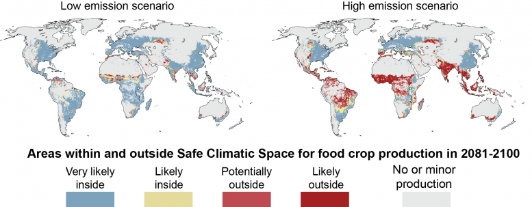
Areas within and outside Safe Climatic Space for food production 2081-2100. Credit: Matti Kummu/Aalto University

New estimates show that if greenhouse gases continue growing at current rates, large regions at risk of being pushed into climate conditions in which no food is grown today.

Climate change is known to negatively affect agriculture and livestock, but there has been little scientific knowledge on which regions of the planet would be touched or what the biggest risks may be. New research led by Aalto University assesses just how global food production will be affected if greenhouse gas emissions are left uncut. The study is published in the prestigious journal One Earth on today (Friday, May 14, 2021).

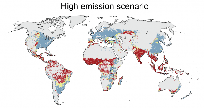
“Our research shows that rapid, out-of-control growth of greenhouse gas emissions may, by the end of the century, lead to more than a third of current global food production falling into conditions in which no food is produced today — that is, out of safe climatic space,” explains Matti Kummu, professor of global water and food issues at Aalto University.

According to the study, this scenario is likely to occur if carbon dioxide emissions continue growing at current rates. In the study, the researchers define the concept of safe climatic space as those areas where 95% of crop production currently takes place, thanks to a combination of three climate factors, rainfall, temperature, and aridity.

Global Food Production High Emissions Scenario

High emissions scenario: areas within and outside Safe Climatic Space for food production 2081-2100 (see comparison image for legend). Credit: Matti Kummu/Aalto University

“The good news is that only a fraction of food production would face as-of-yet unseen conditions if we collectively reduce emissions, so that warming would be limited to 1.5 to 2 degrees Celsius,” says Kummu.

Changes in rainfall and aridity as well as the warming climate are especially threatening to food production in South and Southeast Asia as well as the Sahel region of Africa. These are also areas that lack the capacity to adapt to changing conditions.

“Food production as we know it developed under a fairly stable climate, during a period of slow warming that followed the last ice age. The continuous growth of greenhouse gas emissions may create new conditions, and food crop and livestock production just won’t have enough time to adapt,” says Doctoral Candidate Matias Heino, the other main author of the publication.

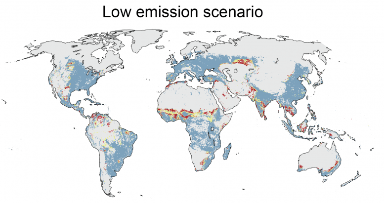
Global Food Production Low Emissions Scenario

Close-up of global low emissions scenario: areas within and outside Safe Climatic Space for food production 2081-2100 (see comparison image for legend). Credit: Matti Kummu/Aalto University

Two future scenarios for climate change were used in the study: one in which carbon dioxide emissions are cut radically, limiting global warming to 1.5-2 degrees Celsius, and another in which emissions continue growing unhalted.

The researchers assessed how climate change would affect 27 of the most important food crops and seven different livestock, accounting for societies’ varying capacities to adapt to changes. The results show that threats affect countries and continents in different ways; in 52 of the 177 countries studied, the entire food production would remain in the safe climatic space in the future. These include Finland and most other European countries.

Already vulnerable countries such as Benin, Cambodia, Ghana, Guinea-Bissau, Guyana and Suriname will be hit hard if no changes are made; up to 95 percent of current food production would fall outside of safe climatic space. Alarmingly, these nations also have significantly less capacity to adapt to changes brought on by climate change when compared to rich Western countries. In all, 20% of the world’s crop production and 18% of livestock production under threat are located in countries with low resilience to adapt to changes.

If carbon dioxide emissions are brought under control, the researchers estimate that the world’s largest climatic zone of today — the boreal forest, which stretches across northern North America, Russia and Europe — would shrink from its current 18.0 to 14.8 million square kilometers by 2100. Should we not be able to cut emissions, only roughly 8 million square kilometers of the vast forest would remain. The change would be even more dramatic in North America: in 2000, the zone covered approximately 6.7 million square kilometers — by 2090 it may shrink to one-third.

Arctic tundra would be even worse off: it is estimated to disappear completely if climate change is not reined in. At the same time, the tropical dry forest and tropical desert zones are estimated to grow.

“If we let emissions grow, the increase in desert areas is especially troubling because in these conditions barely anything can grow without irrigation. By the end of this century, we could see more than 4 million square kilometers of new desert around the globe,” Kummu says.

While the study is the first to take a holistic look at the climatic conditions where food is grown today and how climate change will affect these areas in coming decades, its take-home message is by no means unique: the world needs urgent action.

“We need to mitigate climate change and, at the same time, boost the resilience of our food systems and societies — we cannot leave the vulnerable behind. Food production must be sustainable,” says Heino.

Reference: “Climate change risks pushing one-third of global food production outside the safe climatic space” by Matti Kummu, Matias Heino, Maija Taka, Olli Varis and Daniel Viviroli, 14 May 2021, One Earth.
DOI: 10.1016/j.oneear.2021.04.017

How to Keep Shared Spacesuit
How to Keep Shared Spacesuit “Underwear” Clean?
Spacewalking

Astronaut David A. Wolf, mission specialist, his feet securely planted in a restraint device on the end of the Space Station Remote Manipulator System (SSRMS) or Canadarm2, appears suspended over a heavily cloud-covered part of Earth. Astronauts Wolf and Piers J. Sellers were the assigned spacewalkers for this the second STS-112 spacewalk as well as the two others. Credit: NASA

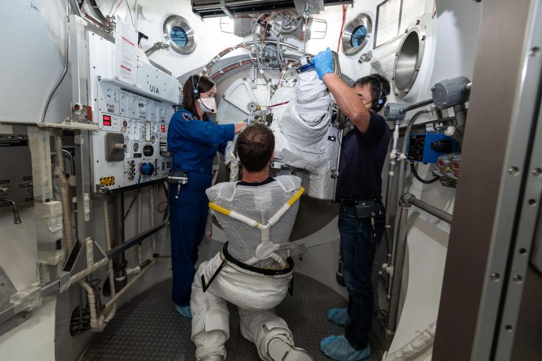
Spacewalking is a major highlight of any astronaut’s career. But there is a downside: putting on your spacesuit means sharing some previously-worn underlayers. A new ESA study is looking into how best to keep these items clean and hygienic as humans venture on to the Moon and beyond.

During the Space Shuttle era, each astronaut was issued with their own ‘External Mobility Unit,’ the official term for a spacesuit. But crews aboard the International Space Station have shifted to sharing suits, with differently sized segments put together to fit a given spacewalker.

Liquid Cooling and Ventilation Garment

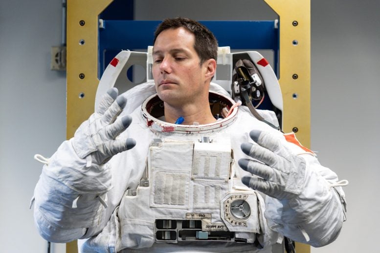
ESA astronaut Thomas Pesquet putting on his External Mobility Unit (EMU) spacesuit, with his Liquid Cooling and Ventilation Garment visible. Thomas donned the spacesuit fit check in the Space Station Airlock Test Article (SSATA) in the Crew Systems Laboratory at NASA’s Johnson Space Center in September 2020, ahead of his Expedition 65 mission to the Intermational Space Station. Credit: NASA-Robert Markowitz

The first item spacewalkers put on is a (disposable) ‘Maximum Absorbency Garment’ diaper, then their own ‘Thermal Comfort Undergarment,’ followed by the long-underwear-like Liquid Cooling and Ventilation Garment (LCVG). Worn next to the skin, the LCVG incorporates liquid cooling tubes and gas ventilation to keep its wearer cool and comfortable during the sustained physical exertion of work in hard vacuum.

But the LCVG is reused by different spacewalkers along with the spacesuits themselves. Such reuse is expected to grow once crews are established aboard the Gateway later this decade, a new international space station in lunar orbit.

With such long-term sharing in mind, ESA has commenced a new project called ‘Biocidal Advanced Coating Technology for Reducing Microbial Activity,’ or BACTeRMA for short.

Trying on Spacesuit

ESA astronaut Thomas Pesquet trying out a spacesuit. The gloves are made to measure and the legs and arms can be adjusted at will, but the torso is rigid and only comes in three sizes: M, L, and XL. This picture was taken at the Sonny Carter Training Facility – Flight Crew Equipment Suit Lab of NASA’s Johnson Space Center. Credit: NASA-Robert Markowitz

“Spaceflight textiles, especially when subject to biological contamination – for example, spacesuit underwear – may pose both engineering and medical risks during long duration flights,” explains ESA material engineer Malgorzata Holynska.

“We are already investigating candidate materials for outer spacesuit layers so this early technology development project is a useful complement, looking into small bacteria-killing molecules that may be useful for all kinds of spaceflight textiles, including spacesuit interiors.”

Gateway With I Hab

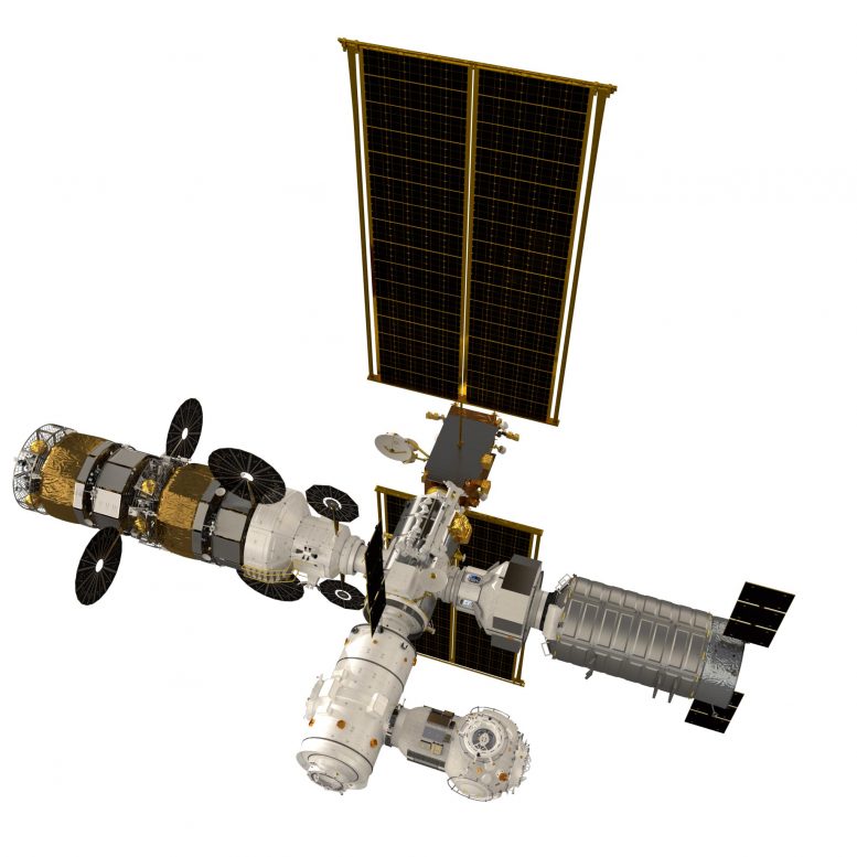
Artist’s impression of the lunar Gateway. Its flight path is a highly-elliptical orbit around the Moon – bringing it both relatively close to the Moon’s surface but also far away making it easier to pick up astronauts and supplies from Earth – around a five-day trip. Credit: ESA

ESA life support specialist Christophe Lasseur adds: “Hygiene is always a concern aboard the International Space Station. Astronauts wear their clothes on alternating days then eventually they are disposed of – burnt up inside reentering spacecraft. But there are some items and surfaces which have to be shared.”

The standard method of preventing biological contamination is the use of antimicrobial materials such as silver or copper, whose ions in the presence of oxygen or water disrupt the normal working of microbial physiology.

Microscopic View of Textiles

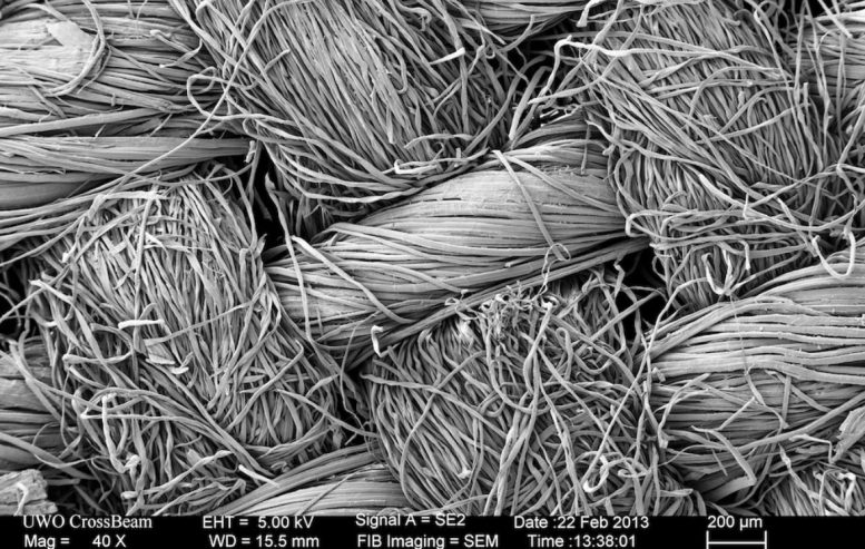
Scanning electron microscope view of test textiles. Credit: OeWF

“The problem is that their long-term use can provoke skin irritation, while the metals themselves may tarnish over time,” explains Seda Özdemir-Fritz  Bacterma project scientist of the Austrian Space Forum (Österreichisches Weltraum Forum /OeWF), the project’s prime contractor.

“To provide an alternative, we are collaborating with the Vienna Textile Lab. They have exclusive access to a unique bacteriographic collection. Those microorganisms produce so-called secondary metabolites. These compounds are typically colorful, and some exhibit versatile properties: antimicrobial, antiviral, and antifungal.

ESA Bacteria

ESA is collaborating with the Austrian Space Forum and the Vienna Textile Lab on using the properties of so-called secondary metabolites to prevent bacterial contamination. Bacterial themselves produce these compounds, which exhibit antimicrobial, antiviral and antifungal properties, to protect themselves from environmental conditions. Credit: OeWF

“It might sound counterintuitive to get rid of microbes using the products of microbes, but all kinds of organisms use secondary metabolites to protect themselves from an extreme environmental conditions. The project will examine them as an innovative antimicrobial textile finish.”

BACTeRMA Project

BACTeRMA Project. Credit: ESA

The project will develop, and test further innovative textile finishes with antimicrobial properties. The Austrian Space Forum together with Vienna Textile Lab will test processed textiles for their antimicrobial properties and will expose them to perspiration and radiation. Simulated lunar dust will also be added to the mix, because the expectation is that the astronauts’ working environment may become dusty after repeated trips to the surface of the Moon or Mars.

“Radiation testing will simulate prolonged storage in the deep space environment,” adds Malgorzata. “Radiation is known to age and degrade textiles in complex ways.”

The idea for the two-year BACTeRMA project was proposed by OeWF in cooperation with the Vienna Textile Lab as subcontractor, through ESA’s Open Space Innovation Platform, seeking out promising ideas for space research from any source.

OeWF is a space research organization: different experts across various science domains come together in the OeWF to work on space topics, with a special focus on spacesuit technology.

“Christopher Columbus needed ship builders to make his journey happen, and that’s the kind of contribution we in the OeWF hope to make,” says Seda Özdemir-Fritz. “We’re interested in the human factors involved in future Moon Mars missions, so we perform ‘analog astronaut’ simulations and analysis.”

POST image The European Times TV
“Oddball Supernova” Appears Strangely Cool Before Exploding – “Stretches What’s Physically Possible!”
Yellow Supergiant in a Close Binary With a Blue, Main Sequence Companion Star

Artist’s impression of a yellow supergiant in a close binary with a blue, main sequence companion star, similar to the properties derived for the 2019yvr progenitor system in Kilpatrick et al. (2021). If the progenitor system to 2019yvr was in such a binary, it must have had a very close interaction that stripped a large amount of hydrogen from the yellow supergiant in the last 100 years before it exploded as a supernova. Credit: Kavli IPMU / Aya Tsuboi

Never-before-seen scenario ‘stretches what’s physically possible.’

A curiously yellow star has caused astrophysicists to reevaluate what’s possible within our universe.

Led by Northwestern University, the international team used NASA’s Hubble Space Telescope to examine the massive star two-and-a-half years before it exploded into a supernova. At the end of their lives, cool, yellow stars are typically shrouded in hydrogen, which conceals the star’s hot, blue interior. But this yellow star, located 35 million lightyears from Earth in the Virgo galaxy cluster, was mysteriously lacking this crucial hydrogen layer at the time of its explosion.

“We haven’t seen this scenario before,” said Northwestern’s Charles Kilpatrick, who led the study. “If a star explodes without hydrogen, it should be extremely blue — really, really hot. It’s almost impossible for a star to be this cool without having hydrogen in its outer layer. We looked at every single stellar model that could explain a star like this, and every single model requires that the star had hydrogen, which, from its supernova, we know it did not. It stretches what’s physically possible.” 

Supernova 2019yvr Explosion Site

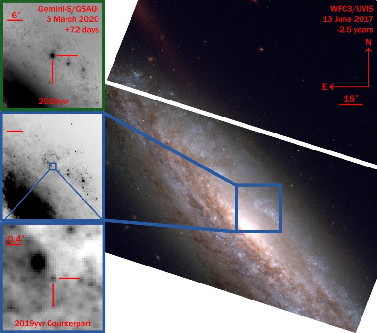
Hubble Space Telescope (HST) imaging showing the explosion site of 2019yvr from 2.5 years before its explosion. Upper left: the supernova itself is seen in an image from the Gemini-South telescope 72 days after it exploded. Lower left: a zoom in to the same site in the pre-explosion HST image, showing a single source that appears to be the progenitor star of 2019yvr. Credit: Charles Kilpatrick / Northwestern University

The team describes the peculiar star and its resulting supernova in a new study, which was published on May 5, 2021, in the Monthly Notices of the Royal Astronomical Society. In the paper, the researchers hypothesize that, in the years preceding its death, the star might have shed its hydrogen layer or lost it to a nearby companion star.

Kilpatrick is a postdoctoral fellow at Northwestern’s Center for Interdisciplinary Exploration and Research in Astrophysics (CIERA) and member of the Young Supernova Experiment, which uses the Pan-STARSS telescope at Haleakalā, Hawaii, to catch supernovae right after they explode. 

Catching a star before it explodes 

After the Young Supernova Experiment spotted supernova 2019yvr in the relatively nearby spiral galaxy NGC 4666, the team used deep space images captured by NASA’s Hubble Space Telescope, which fortunately already observed this section of the sky. 

“What massive stars do right before they explode is a big unsolved mystery,” Kilpatrick said. “It’s rare to see this kind of star right before it explodes into a supernova.”

The Hubble images showed the source of the supernova, a massive star imaged just a couple years before the explosion. Although the supernova itself appeared completely normal, its source — or progenitor star — was anything but.

“When it exploded, it seemed like a very normal hydrogen-free supernova,” Kilpatrick said. “There was nothing outstanding about this. But the progenitor star didn’t match what we know about this type of supernova.” 

Direct evidence of violent death

Several months after the explosion, however, Kilpatrick and his team discovered a clue. As ejecta from the star’s final explosion traveled through its environment, it collided with a large mass of hydrogen. This led the team to hypothesize that the progenitor star might have expelled the hydrogen within a few years before its death. 

“Astronomers have suspected that stars undergo violent eruptions or death throes in the years before we see supernovae,” Kilpatrick said. “This star’s discovery provides some of the most direct evidence ever found that stars experience catastrophic eruptions, which cause them to lose mass before an explosion. If the star was having these eruptions, then it likely expelled its hydrogen several decades before it exploded.”

“This star’s discovery provides some of the most direct evidence ever found that stars experience catastrophic eruptions, which cause them to lose mass before an explosion.”
Charles Kilpatrick, astrophysicist

In the new study, Kilpatrick’s team also presents another possibility: A less massive companion star might have stripped away hydrogen from the supernova’s progenitor star. The team, however, will not be able to search for the companion star until after the supernova’s brightness fades, which could take up to 10 years.

“Unlike it’s normal behavior right after it exploded, the hydrogen interaction revealed it’s kind of this oddball supernova,” Kilpatrick said. “But it’s exceptional that we were able to find its progenitor star in Hubble data. In four or five years, I think we will be able to learn more about what happened.”

For more on this study, read Mysterious Hydrogen-Free Supernova Sheds Light on Massive Stars’ Violent Death Throes.

Reference: “A cool and inflated progenitor candidate for the Type Ib supernova 2019yvr at 2.6 yr before explosion” by Charles D Kilpatrick, Maria R Drout, Katie Auchettl, Georgios Dimitriadis, Ryan J Foley, David O Jones, Lindsay DeMarchi, K Decker French, Christa Gall, Jens Hjorth, Wynn V Jacobson-Galán, Raffaella Margutti, Anthony L Piro, Enrico Ramirez-Ruiz, Armin Rest and César Rojas-Bravo, 30 March 2021, Monthly Notices of the Royal Astronomical Society.
DOI: 10.1093/mnras/stab838

The study, “A cool and inflated progenitor candidate for the type Ib supernova 2019 yvr at 2.6 years before explosion,” was supported by NASA (award numbers GO-15691 and AR-16136), the National Science Foundation (award numbers AST-1909796, AST-1944985), the Canadian Institute for Advanced Research, the VILLUM Foundation and the Australian Research Council Centre of Excellence. In addition to the Hubble Space Telescope, the researchers used instruments at the Gemini Observatory, Keck Observatory, Las Cumbres Observatory, Spitzer Space Telescope and the Swope Telescope.

Scientists Invent New Method for Producing Synthetic DNA

PhD Alexander Sandahl, together with Professor Kurt Gothelf, Professor Troels Skrydstrup and a number of students in the groups, has developed a method for efficient and automated production of ingredients for DNA synthesis. Credit: Colourbox

Chemically synthesized short DNA sequences are extremely important ingredients with countless uses in research laboratories, hospitals, and in industry, like in the method for identifying COVID-19. Phosphoramidites are necessary building blocks in the production of DNA sequences, but they are unstable, and break quickly. PhD Alexander Sandahl (Professor Kurt Gothelf’s group) has, in collaboration with a researcher in Professor Troels Skrydstrup’s group, developed a new patented way to quickly and efficiently manufacture the unstable building blocks immediately before they are to be used, and thus streamline DNA production.

The DNA sequences produced are also called oligonucleotides. These are widely used for disease identification, for the manufacture of oligonucleotide-based drugs, and for several other medical and biotechnological applications. 

The high demand for oligonucleotides therefore requires an efficient automated method for their chemical production. This process relies on phosphoramidites, which are chemical compounds that have the disadvantage of being unstable unless stored at the ideal -20 degrees Celsius.

Instruments used for DNA synthesis are not able to cool down the phosphoramidites, and consequently it is unavoidable that some of them degrade after being added to the instrument. 

Schematic overview of strategy for synthesis of phosphoramidites in a flow-based setup. Credit: Nature Commun 12, Artikel nr. 2760 (2021)

Avoiding unwanted degradation of important ingredients

Professor Kurt Gothelf and Professor Troels Skrydstrup are each heading a research group in organic chemistry, which have worked together to develop a relatively simple but efficient technology where the production of phosphoramidites can be automated and integrated directly into the instrument for DNA synthesis.

This avoids both the manual synthesis of these, which normally would take up to 12 hours, as well as the problem of storing unstable phosphoramidites. Gothelf’s group has contributed with their expertise in automated DNA synthesis and Skrydstrup’s group has contributed with their know-how with chemical reactions that take place in continuously flowing liquids (flow chemistry).

“It has been a very rewarding collaboration which is precisely one of the core values of iNANO,” says Kurt Gothelf, who adds “and I would also like to attribute to Alexander Sandahl a large part of the credit for this project being successful, as he has established the collaboration and has developed and realized a large part of the ideas for the project.” 

The results have just been published in the journal, Nature Communications. 

In the method of producing phosphoramidites, nucleosides (starting materials) are flushed through a solid material (resin), which can potentially be fully integrated into an automated process in the instrument for DNA synthesis. The resin ensures that the nucleosides are rapidly phosphorylated, whereby the nucleosides are converted to phosphoramidites within a few minutes. From the resin, the phosphoramidites are automatically flushed on to the part of the instrument which is responsible for the DNA synthesis.

This avoids the degradation of the phosphoramidites, as they are first produced just before they are to be used (on-demand), in a faster, more efficient flow-based way that can potentially be automated and operated by non-chemists.

Reference: “On-demand synthesis of phosphoramidite” by Alexander F. Sandahl, Thuy J. D. Nguyen, Rikke A. Hansen, Martin B. Johansen, Troels Skrydstrup and Kurt V. Gothelf, 12 May 2021, Nature Communications.
DOI: 10.1038/s41467-021-22945-z

The research is financially supported by the Lundbeck Foundation, the Novo Nordisk Foundation (CEMBID), the Independent Research Fund Denmark, and the Danish National Research Foundation (CADIAC).

Researchers Identify Genes Associated With Significant Increase in COVID-19 Risk

Having genetic risk variants in the ABO gene might significantly increase the chances of developing COVID-19, and other genes may also increase COVID-19 risk, according to research presented at the ATS 2021 International Conference.

Much about COVID-19 remains a medical mystery, including whether certain genes place individuals at greater risk of contracting the SARS-CoV-2 virus, which causes COVID-19. Ana Hernandez Cordero, PhD, postdoctoral fellow with the Centre for Heart Lung Innovation, University of British Columbia, and colleagues used integrative genomics combined with proteomics to identify these genes.

Genomic research identifies specific genes that may play a role in biological processes such as the development of disease, while proteomics does the same for proteins. Researchers can get a fuller picture of disease processes by integrating tools to investigate both.

DNA is a big, complex molecule and so, genetic associations alone cannot pinpoint the exact gene responsible for COVID-19,” said Dr. Hernandez. “However, by combining COVID-19 genetic information with gene expression and proteomic datasets, we can figure out which genes are driving the relationship with COVID-19.”

The researchers combined genetic information with an examination of lung gene expression to identify genetic variants that were controlling gene expression in the lung that were responsible for COVID-19. The researchers identified specific genes’ markers that share their effects on gene expression and protein levels with COVID-19 susceptibility. For the analysis, they used bioinformatics to integrate: (1) a genomic dataset obtained from patients who were infected with SARS-CoV-2 as well as non-infected individuals (controls); (2) lung and blood tissue gene expression datasets from clinical populations (non-COVID-19); and (3) a proteome dataset obtained from blood donors (non-COVID-19).

By doing this, they found that several genes responsible for the immune system’s response to COVID-19 are also involved in COVID-19 susceptibility. What they discovered was supported by the findings of previous research.

Looking for candidate genes in blood proteins, they were able to go one step further in connecting the effects of genes to susceptibility to COVID-19. Blood proteomics can also help identify markers in the blood that can be easily measured to indicate disease status, and potentially, to monitor the disease.

“By harnessing the power of genomic information, we identified genes that are related to COVID-19,” said Dr. Hernandez. “In particular, we found that the ABO gene is a significant risk factor for COVID-19. Of particular note was the relationship between the blood group ABO and COVID-19 risk. We showed that the relationship is not just an association but causal.”

In addition to the ABO gene, Dr. Hernandez and colleagues found that people carrying certain genetic variants for SLC6A20, ERMP1, FCER1G and CA11 have a significantly higher risk of contracting COVID-19. “These individuals should use extreme caution during the pandemic. These genes may also prove to be good markers for disease as well as potential drug targets.”

Several of the genes identified in the researchers’ analysis have already been linked with respiratory diseases. For example, ERMP1 has been linked to asthma. CA11 may also elevate COVID-19 risk for people with diabetes.

Genetic associations for COVID-19 and gene and protein expression were combined using integrative genomics (IG). IG aims to identify mechanisms (for example: gene expression levels) that connect the effects of the genetic code to a complex disease. These methods, although complex, are also fast and their outcomes can help researchers to prioritize candidate genes for in vitro (in the lab) and in vivo (in living organisms) testing.

Dr. Hernandez added, “Our research has progressed since the time that we first conducted this analysis. We have now identified even more interesting candidates for COVID-19 such as IL10RB, IFNAR2 and OAS1. These genes have been linked to severe COVID-19. Their role in the immune response to viral infections and mounting evidence suggest that these candidates and their role in COVID-19 should be further investigated.”

Reference: “Integrative Genomic Analysis Highlights Potential Genetic Risk Factors for Covid-19” by A. I. Hernandez Cordero, X. Li1, S. Milne, C. Yang, Y. Bossé, P. Joubert, W. Timens, M. Van den Berge, D. Nickle, K. Hao and D. D. Sin, 3 May 2021, ATS 2021 International Conference.
Abstract

New Immunotherapy
New Immunotherapy “Highly Effective” Against Hepatitis B Virus

Immunotherapy Concept

Scientists at University College London have identified a new immunotherapy to combat the hepatitis B virus (HBV), the most common cause of liver cancer in the world.

Each year, globally, chronic HBV causes an estimated 880,000 deaths from liver cirrhosis and hepatocellular carcinoma/liver cancer (HCC).

The pioneering study used immune cells isolated directly from patient liver and tumour tissue, to show that targeting acyl-CoA:cholesterol acyltransferase (ACAT), an enzyme that helps to manage cholesterol levels in cells*, was highly effective at boosting immune responses.

Published in Nature Communications, the findings show that blocking the activity of ACAT with ACAT inhibitors boosts the specific immune cells that can fight both the virus and associated cancerous tumours, demonstrating its effectiveness as an immunotherapy. Inhibiting ACAT was also found to impede HBV’s own replication, thereby also acting as a direct antiviral. ACAT inhibitors such as avasimibe, taken orally, have previously been shown to be well-tolerated as cholesterol-lowering drugs in humans.

Explaining the study, lead author Professor Mala Maini (UCL Division of Infection & Immunity), said: “Chronic hepatitis B virus infection is a major global health problem and the most common cause of liver cancer in the world.

“The development of novel therapeutic options is crucial to improve patient care. Immune cells such as T cells are indispensable for fighting viruses and tumours but are often highly dysfunctional and fail to control these diseases. Current standard of care treatments are often incapable of eliminating the virus, do not prevent cancer development and do not rescue immune cells.

“In this study, we aimed to identify a treatment target to directly inhibit the virus while also boosting the immune cells fighting it.”

Cholesterol is a lipid (fat) that we ingest every day in our diets and that can exert multiple functions within different cells of the body. HBV infects the liver, an organ highly enriched in cholesterol and well known for limiting local immune responses.

In this study, using human liver disease tissue samples in vitro, Professor Maini’s lab at UCL showed that ACAT inhibitors boosted human antiviral T cells capable of eliminating the virus. This response is in contrast to currently available therapies. The immune-boosting effect was especially striking in T cells found in the HBV-infected liver and within liver cancer, overcoming the local restraints on immune cell function, allowing the T cells to target both the virus and cancerous cells.

The Maini group then collaborated with Professor Jane McKeating’s lab at the University of Oxford to show that ACAT inhibitors could also block the HBV life cycle in a way that other antivirals are unable to. These drugs therefore have a unique combination of antiviral and immunotherapeutic effects.

Commenting on the findings, first author Dr. Nathalie Schmidt (UCL Division of Infection & Immunity), said: “We have found a highly effective novel target for the treatment of chronic hepatitis B virus infection and liver cancer.

“Modulating cholesterol metabolism with ACAT inhibitors has the unique features of directly targeting the virus and tumours while at the same time boosting the T cells that fight them. This enables us to tackle the disease from multiple directions at the same time.”

Dr. Schmidt added: “The cholesterol-modifying drug is already known to be safe in humans and we hope that our study now informs the development of clinical trials combining cholesterol modulation with other immunotherapies. In summary, our findings offer exciting new possibilities for the treatment of patients with chronic viral infections and cancer.”

*The enzyme is required for cholesterol esterification, a mechanism which prevents excessive cellular levels of cholesterol, which can be toxic to cells.

Reference: 14 May 2021, Nature Communications.
DOI: 10.1038/s41467-021-22967-7

This research was carried out by researchers at UCL, supported by the University of Oxford, the Royal Free London NHS Trust, and Leiden University Medical Centre, the Netherlands.

Grant funding came from the Wellcome Trust and Cancer Research UK.

Researchers Develop 3D-Printed Jelly for Biomedical Materials and Soft Robotics
3D-Printed Jelly

The hydrogel material comes from different-sized seaweed particles. Credit: Orlin Velev, NC State University

Hydrogels merge two physical forms of the same seaweed material for strength, flexibility.

3D-printable gels with improved and highly controlled properties can be created by merging micro- and nano-sized networks of the same materials harnessed from seaweed, according to new research from North Carolina State University. The findings could have applications in biomedical materials — think of biological scaffolds for growing cells — and soft robotics.

Described in the journal Nature Communications, the findings show that these water-based gels — called homocomposite hydrogels — are both strong and flexible. They are composed of alginates — chemical compounds found in seaweed and algae that are commonly used as thickening agents and in wound dressings.

Merging different-size scale networks of the same alginate together eliminates the fragility that can sometimes occur when differing materials are merged together in a hydrogel, says Orlin Velev, S. Frank and Doris Culberson Distinguished Professor of Chemical and Biomolecular Engineering at NC State and corresponding author of the paper.

“Water-based materials can be soft and brittle,” he said. “But these homocomposite materials — soft fibrillar alginate particles inside a medium of alginate — are really two hydrogels in one: one is a particle hydrogel and one is a molecular hydrogel. Merged together they produce a jelly-like material that is better than the sum of its parts, and whose properties can be tuned precisely for shaping through a 3D printer for on-demand manufacturing.”

“We are reinforcing a hydrogel material with the same material, which is remarkable because it uses just one material to improve the overall mechanical properties,” said Lilian Hsiao, an assistant professor of chemical and molecular engineering at NC State and a co-author of the paper. “Alginates are used in wound dressings, so this material potentially could be used as a strengthened 3D-printed bandage or as a patch for wound healing or drug delivery.”

“These types of materials have the potential to be most useful in medical products, in food products as a thickening agent, or in soft robotics,” said Austin Williams, one of the paper’s first coauthors and a graduate student in Velev’s lab.

Future work will attempt to fine-tune this method of merging of homocomposite materials to advance 3D printing for biomedical applications or biomedical injection materials, Velev said.

“This technique may have uses with other types of gels, like those used in coatings or in consumer products,” Hsiao said.

Reference: “Printable homocomposite hydrogels with synergistically reinforced molecular-colloidal networks” by Austin Williams, Sangchul Roh, Alan Jacob, Lilian Hsiao, Orlin D. Velev and Simeon Stoyanov, 14 May 2021, Nature Communications.
DOI: 10.1038/s41467-021-23098-9

Former NC State Ph.D. student Sangchul Roh is the paper’s other first coauthor. Coauthor Simeon Stoyanov from Wageningen University participated in the conception of the new material.

The research is funded by the National Science Foundation under grants CMMI-1825476, CBET-1804462 and ECCS-2025064.

Watch NASA's Ingenuity Mars Helicopter Fly in 3D
Watch NASA’s Ingenuity Mars Helicopter Fly in 3D

A new video of the helicopter’s third flight gives viewers the sensation of standing on the Red Planet and seeing the action firsthand.

When NASA’s Ingenuity Mars Helicopter took to the Martian skies on its third flight on April 25, the agency’s Perseverance rover was there to capture the historic moment. Now NASA engineers have rendered the flight in 3D, lending dramatic depth to the flight as the helicopter ascends, hovers, then zooms laterally off-screen before returning for a pinpoint landing. Seeing the sequence is a bit like standing on the Martian surface next to Perseverance and watching the flight firsthand.

Located on the rover’s mast, or “head,” the zoomable dual-camera Mastcam-Z imager provided the view. Along with producing images that enable the public to follow the rover’s daily discoveries, the cameras provide key data to help engineers navigate and scientists choose interesting rocks to study.

Ingenuity Flies in 3D: After the Mastcam-Z imager aboard NASA’s Perseverance rover captured Ingenuity’s third flight on April 25, the frames of the video that was stitched together were then reprojected to optimize viewing in an anaglyph, or an image seen in 3D when seen through color-filtered glasses. Credit: NASA/JPL-Caltech/ASU/MSSS

Justin Maki, an imaging scientist at NASA’s Jet Propulsion Laboratory in Southern California, led the team that stitched the images into a video. The frames of the video were reprojected to optimize viewing in an anaglyph, or an image seen in 3D when viewed with color-filtered glasses (you can create your own 3D glasses in a few minutes).

Maki’s been creating 3D imaging of Mars since his days as a graduate student processing images from NASA’s Sojourner, the first Mars rover in 1997. But this is the first time he’s created actual 3D video of an aircraft flying on Mars. “The Mastcam-Z video capability was inherited from the Mars Science Laboratory MARDI (MArs Descent Imager) camera,” Maki said. “To be reusing this capability on a new mission by acquiring 3D video of a helicopter flying above the surface of Mars is just spectacular.” The videos of the helicopter are the most extensive 3D video yet from the Mastcam-Z team.

The rover’s drivers and robotic-arm operators use a more sophisticated 3D system to understand exactly how things are positioned on Mars before planning the rover’s movements. But, according to Maki, team members have also been viewing still 3D images for rover-drive planning. “A helicopter flying on Mars opens a new era for Mars exploration. It’s a great demonstration of a new technology for exploration,” he added. “With each flight we open up more possibilities.”

Perseverance Rover’s Mastcam-Z Captures Ingenuity’s Third Flight: NASA’s Ingenuity Mars Helicopter takes off and lands in this video captured on April 25, 2021, by Mastcam-Z, an imager aboard NASA’s Perseverance Mars rover. As expected, the helicopter flew out of its field of vision while completing a flight plan that took it 164 feet (50 meters) downrange of the landing spot. Keep watching, the helicopter will return to stick the landing. Credits: NASA/JPL-Caltech/ASU/MSSS

The April 25 flight brought with it several other firsts, with Ingenuity rising 16 feet (5 meters), then flying downrange 164 feet (50 meters). That was a record until Ingenuity traveled 873 feet (266 meters) on its fourth flight, on April 30. For its fifth flight, on May 7, Ingenuity completed its first one-way trip, traveling 423 feet (129 meters), then reaching an altitude of 33 feet (10 meters) above its new landing field.

The flights began as a technology demonstration intended to prove that powered, controlled flight on Mars is possible. Now they will serve as an operations demonstration, exploring how aerial scouting and other functions could benefit future exploration of Mars.