How to Keep Shared Spacesuit
How to Keep Shared Spacesuit “Underwear” Clean?
Spacewalking

Astronaut David A. Wolf, mission specialist, his feet securely planted in a restraint device on the end of the Space Station Remote Manipulator System (SSRMS) or Canadarm2, appears suspended over a heavily cloud-covered part of Earth. Astronauts Wolf and Piers J. Sellers were the assigned spacewalkers for this the second STS-112 spacewalk as well as the two others. Credit: NASA

Spacewalking is a major highlight of any astronaut’s career. But there is a downside: putting on your spacesuit means sharing some previously-worn underlayers. A new ESA study is looking into how best to keep these items clean and hygienic as humans venture on to the Moon and beyond.

During the Space Shuttle era, each astronaut was issued with their own ‘External Mobility Unit,’ the official term for a spacesuit. But crews aboard the International Space Station have shifted to sharing suits, with differently sized segments put together to fit a given spacewalker.
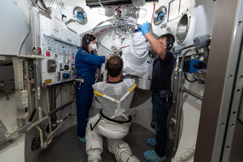
Liquid Cooling and Ventilation Garment

ESA astronaut Thomas Pesquet putting on his External Mobility Unit (EMU) spacesuit, with his Liquid Cooling and Ventilation Garment visible. Thomas donned the spacesuit fit check in the Space Station Airlock Test Article (SSATA) in the Crew Systems Laboratory at NASA’s Johnson Space Center in September 2020, ahead of his Expedition 65 mission to the Intermational Space Station. Credit: NASA-Robert Markowitz

The first item spacewalkers put on is a (disposable) ‘Maximum Absorbency Garment’ diaper, then their own ‘Thermal Comfort Undergarment,’ followed by the long-underwear-like Liquid Cooling and Ventilation Garment (LCVG). Worn next to the skin, the LCVG incorporates liquid cooling tubes and gas ventilation to keep its wearer cool and comfortable during the sustained physical exertion of work in hard vacuum.

But the LCVG is reused by different spacewalkers along with the spacesuits themselves. Such reuse is expected to grow once crews are established aboard the Gateway later this decade, a new international space station in lunar orbit.

With such long-term sharing in mind, ESA has commenced a new project called ‘Biocidal Advanced Coating Technology for Reducing Microbial Activity,’ or BACTeRMA for short.
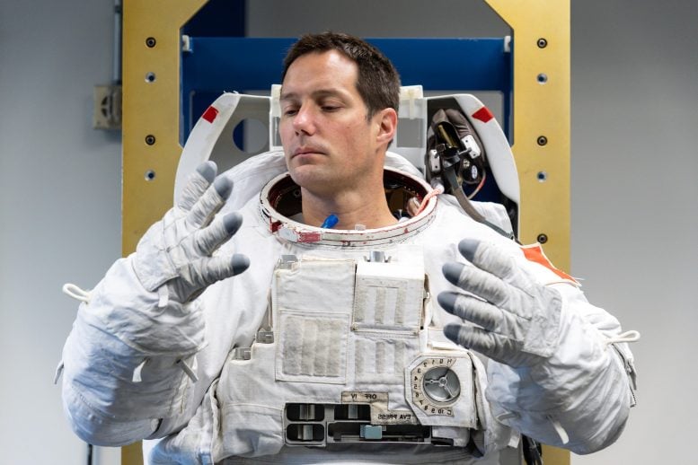
Trying on Spacesuit

ESA astronaut Thomas Pesquet trying out a spacesuit. The gloves are made to measure and the legs and arms can be adjusted at will, but the torso is rigid and only comes in three sizes: M, L, and XL. This picture was taken at the Sonny Carter Training Facility – Flight Crew Equipment Suit Lab of NASA’s Johnson Space Center. Credit: NASA-Robert Markowitz

“Spaceflight textiles, especially when subject to biological contamination – for example, spacesuit underwear – may pose both engineering and medical risks during long duration flights,” explains ESA material engineer Malgorzata Holynska.

“We are already investigating candidate materials for outer spacesuit layers so this early technology development project is a useful complement, looking into small bacteria-killing molecules that may be useful for all kinds of spaceflight textiles, including spacesuit interiors.”
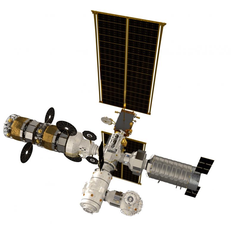
Gateway With I Hab

Artist’s impression of the lunar Gateway. Its flight path is a highly-elliptical orbit around the Moon – bringing it both relatively close to the Moon’s surface but also far away making it easier to pick up astronauts and supplies from Earth – around a five-day trip. Credit: ESA

ESA life support specialist Christophe Lasseur adds: “Hygiene is always a concern aboard the International Space Station. Astronauts wear their clothes on alternating days then eventually they are disposed of – burnt up inside reentering spacecraft. But there are some items and surfaces which have to be shared.”

The standard method of preventing biological contamination is the use of antimicrobial materials such as silver or copper, whose ions in the presence of oxygen or water disrupt the normal working of microbial physiology.

Microscopic View of Textiles

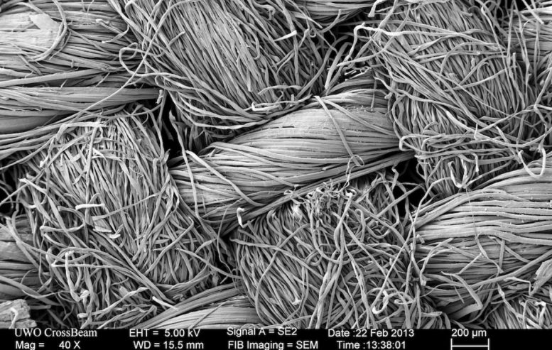
Scanning electron microscope view of test textiles. Credit: OeWF

“The problem is that their long-term use can provoke skin irritation, while the metals themselves may tarnish over time,” explains Seda Özdemir-Fritz  Bacterma project scientist of the Austrian Space Forum (Österreichisches Weltraum Forum /OeWF), the project’s prime contractor.

“To provide an alternative, we are collaborating with the Vienna Textile Lab. They have exclusive access to a unique bacteriographic collection. Those microorganisms produce so-called secondary metabolites. These compounds are typically colorful, and some exhibit versatile properties: antimicrobial, antiviral, and antifungal.

ESA Bacteria

ESA is collaborating with the Austrian Space Forum and the Vienna Textile Lab on using the properties of so-called secondary metabolites to prevent bacterial contamination. Bacterial themselves produce these compounds, which exhibit antimicrobial, antiviral and antifungal properties, to protect themselves from environmental conditions. Credit: OeWF

“It might sound counterintuitive to get rid of microbes using the products of microbes, but all kinds of organisms use secondary metabolites to protect themselves from an extreme environmental conditions. The project will examine them as an innovative antimicrobial textile finish.”

BACTeRMA Project

BACTeRMA Project. Credit: ESA

The project will develop, and test further innovative textile finishes with antimicrobial properties. The Austrian Space Forum together with Vienna Textile Lab will test processed textiles for their antimicrobial properties and will expose them to perspiration and radiation. Simulated lunar dust will also be added to the mix, because the expectation is that the astronauts’ working environment may become dusty after repeated trips to the surface of the Moon or Mars.

“Radiation testing will simulate prolonged storage in the deep space environment,” adds Malgorzata. “Radiation is known to age and degrade textiles in complex ways.”

The idea for the two-year BACTeRMA project was proposed by OeWF in cooperation with the Vienna Textile Lab as subcontractor, through ESA’s Open Space Innovation Platform, seeking out promising ideas for space research from any source.

OeWF is a space research organization: different experts across various science domains come together in the OeWF to work on space topics, with a special focus on spacesuit technology.

“Christopher Columbus needed ship builders to make his journey happen, and that’s the kind of contribution we in the OeWF hope to make,” says Seda Özdemir-Fritz. “We’re interested in the human factors involved in future Moon Mars missions, so we perform ‘analog astronaut’ simulations and analysis.”

POST image The European Times TV
“Oddball Supernova” Appears Strangely Cool Before Exploding – “Stretches What’s Physically Possible!”
Yellow Supergiant in a Close Binary With a Blue, Main Sequence Companion Star

Artist’s impression of a yellow supergiant in a close binary with a blue, main sequence companion star, similar to the properties derived for the 2019yvr progenitor system in Kilpatrick et al. (2021). If the progenitor system to 2019yvr was in such a binary, it must have had a very close interaction that stripped a large amount of hydrogen from the yellow supergiant in the last 100 years before it exploded as a supernova. Credit: Kavli IPMU / Aya Tsuboi

Never-before-seen scenario ‘stretches what’s physically possible.’

A curiously yellow star has caused astrophysicists to reevaluate what’s possible within our universe.

Led by Northwestern University, the international team used NASA’s Hubble Space Telescope to examine the massive star two-and-a-half years before it exploded into a supernova. At the end of their lives, cool, yellow stars are typically shrouded in hydrogen, which conceals the star’s hot, blue interior. But this yellow star, located 35 million lightyears from Earth in the Virgo galaxy cluster, was mysteriously lacking this crucial hydrogen layer at the time of its explosion.

“We haven’t seen this scenario before,” said Northwestern’s Charles Kilpatrick, who led the study. “If a star explodes without hydrogen, it should be extremely blue — really, really hot. It’s almost impossible for a star to be this cool without having hydrogen in its outer layer. We looked at every single stellar model that could explain a star like this, and every single model requires that the star had hydrogen, which, from its supernova, we know it did not. It stretches what’s physically possible.” 

Supernova 2019yvr Explosion Site

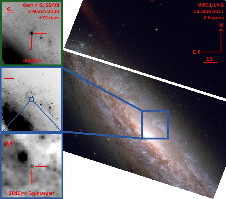
Hubble Space Telescope (HST) imaging showing the explosion site of 2019yvr from 2.5 years before its explosion. Upper left: the supernova itself is seen in an image from the Gemini-South telescope 72 days after it exploded. Lower left: a zoom in to the same site in the pre-explosion HST image, showing a single source that appears to be the progenitor star of 2019yvr. Credit: Charles Kilpatrick / Northwestern University

The team describes the peculiar star and its resulting supernova in a new study, which was published on May 5, 2021, in the Monthly Notices of the Royal Astronomical Society. In the paper, the researchers hypothesize that, in the years preceding its death, the star might have shed its hydrogen layer or lost it to a nearby companion star.

Kilpatrick is a postdoctoral fellow at Northwestern’s Center for Interdisciplinary Exploration and Research in Astrophysics (CIERA) and member of the Young Supernova Experiment, which uses the Pan-STARSS telescope at Haleakalā, Hawaii, to catch supernovae right after they explode. 

Catching a star before it explodes 

After the Young Supernova Experiment spotted supernova 2019yvr in the relatively nearby spiral galaxy NGC 4666, the team used deep space images captured by NASA’s Hubble Space Telescope, which fortunately already observed this section of the sky. 

“What massive stars do right before they explode is a big unsolved mystery,” Kilpatrick said. “It’s rare to see this kind of star right before it explodes into a supernova.”

The Hubble images showed the source of the supernova, a massive star imaged just a couple years before the explosion. Although the supernova itself appeared completely normal, its source — or progenitor star — was anything but.

“When it exploded, it seemed like a very normal hydrogen-free supernova,” Kilpatrick said. “There was nothing outstanding about this. But the progenitor star didn’t match what we know about this type of supernova.” 

Direct evidence of violent death

Several months after the explosion, however, Kilpatrick and his team discovered a clue. As ejecta from the star’s final explosion traveled through its environment, it collided with a large mass of hydrogen. This led the team to hypothesize that the progenitor star might have expelled the hydrogen within a few years before its death. 

“Astronomers have suspected that stars undergo violent eruptions or death throes in the years before we see supernovae,” Kilpatrick said. “This star’s discovery provides some of the most direct evidence ever found that stars experience catastrophic eruptions, which cause them to lose mass before an explosion. If the star was having these eruptions, then it likely expelled its hydrogen several decades before it exploded.”

“This star’s discovery provides some of the most direct evidence ever found that stars experience catastrophic eruptions, which cause them to lose mass before an explosion.”
Charles Kilpatrick, astrophysicist

In the new study, Kilpatrick’s team also presents another possibility: A less massive companion star might have stripped away hydrogen from the supernova’s progenitor star. The team, however, will not be able to search for the companion star until after the supernova’s brightness fades, which could take up to 10 years.

“Unlike it’s normal behavior right after it exploded, the hydrogen interaction revealed it’s kind of this oddball supernova,” Kilpatrick said. “But it’s exceptional that we were able to find its progenitor star in Hubble data. In four or five years, I think we will be able to learn more about what happened.”

For more on this study, read Mysterious Hydrogen-Free Supernova Sheds Light on Massive Stars’ Violent Death Throes.

Reference: “A cool and inflated progenitor candidate for the Type Ib supernova 2019yvr at 2.6 yr before explosion” by Charles D Kilpatrick, Maria R Drout, Katie Auchettl, Georgios Dimitriadis, Ryan J Foley, David O Jones, Lindsay DeMarchi, K Decker French, Christa Gall, Jens Hjorth, Wynn V Jacobson-Galán, Raffaella Margutti, Anthony L Piro, Enrico Ramirez-Ruiz, Armin Rest and César Rojas-Bravo, 30 March 2021, Monthly Notices of the Royal Astronomical Society.
DOI: 10.1093/mnras/stab838

The study, “A cool and inflated progenitor candidate for the type Ib supernova 2019 yvr at 2.6 years before explosion,” was supported by NASA (award numbers GO-15691 and AR-16136), the National Science Foundation (award numbers AST-1909796, AST-1944985), the Canadian Institute for Advanced Research, the VILLUM Foundation and the Australian Research Council Centre of Excellence. In addition to the Hubble Space Telescope, the researchers used instruments at the Gemini Observatory, Keck Observatory, Las Cumbres Observatory, Spitzer Space Telescope and the Swope Telescope.

Scientists Invent New Method for Producing Synthetic DNA

PhD Alexander Sandahl, together with Professor Kurt Gothelf, Professor Troels Skrydstrup and a number of students in the groups, has developed a method for efficient and automated production of ingredients for DNA synthesis. Credit: Colourbox

Chemically synthesized short DNA sequences are extremely important ingredients with countless uses in research laboratories, hospitals, and in industry, like in the method for identifying COVID-19. Phosphoramidites are necessary building blocks in the production of DNA sequences, but they are unstable, and break quickly. PhD Alexander Sandahl (Professor Kurt Gothelf’s group) has, in collaboration with a researcher in Professor Troels Skrydstrup’s group, developed a new patented way to quickly and efficiently manufacture the unstable building blocks immediately before they are to be used, and thus streamline DNA production.

The DNA sequences produced are also called oligonucleotides. These are widely used for disease identification, for the manufacture of oligonucleotide-based drugs, and for several other medical and biotechnological applications. 

The high demand for oligonucleotides therefore requires an efficient automated method for their chemical production. This process relies on phosphoramidites, which are chemical compounds that have the disadvantage of being unstable unless stored at the ideal -20 degrees Celsius.

Instruments used for DNA synthesis are not able to cool down the phosphoramidites, and consequently it is unavoidable that some of them degrade after being added to the instrument. 

Schematic overview of strategy for synthesis of phosphoramidites in a flow-based setup. Credit: Nature Commun 12, Artikel nr. 2760 (2021)

Avoiding unwanted degradation of important ingredients

Professor Kurt Gothelf and Professor Troels Skrydstrup are each heading a research group in organic chemistry, which have worked together to develop a relatively simple but efficient technology where the production of phosphoramidites can be automated and integrated directly into the instrument for DNA synthesis.

This avoids both the manual synthesis of these, which normally would take up to 12 hours, as well as the problem of storing unstable phosphoramidites. Gothelf’s group has contributed with their expertise in automated DNA synthesis and Skrydstrup’s group has contributed with their know-how with chemical reactions that take place in continuously flowing liquids (flow chemistry).

“It has been a very rewarding collaboration which is precisely one of the core values of iNANO,” says Kurt Gothelf, who adds “and I would also like to attribute to Alexander Sandahl a large part of the credit for this project being successful, as he has established the collaboration and has developed and realized a large part of the ideas for the project.” 

The results have just been published in the journal, Nature Communications. 

In the method of producing phosphoramidites, nucleosides (starting materials) are flushed through a solid material (resin), which can potentially be fully integrated into an automated process in the instrument for DNA synthesis. The resin ensures that the nucleosides are rapidly phosphorylated, whereby the nucleosides are converted to phosphoramidites within a few minutes. From the resin, the phosphoramidites are automatically flushed on to the part of the instrument which is responsible for the DNA synthesis.

This avoids the degradation of the phosphoramidites, as they are first produced just before they are to be used (on-demand), in a faster, more efficient flow-based way that can potentially be automated and operated by non-chemists.

Reference: “On-demand synthesis of phosphoramidite” by Alexander F. Sandahl, Thuy J. D. Nguyen, Rikke A. Hansen, Martin B. Johansen, Troels Skrydstrup and Kurt V. Gothelf, 12 May 2021, Nature Communications.
DOI: 10.1038/s41467-021-22945-z

The research is financially supported by the Lundbeck Foundation, the Novo Nordisk Foundation (CEMBID), the Independent Research Fund Denmark, and the Danish National Research Foundation (CADIAC).

Researchers Identify Genes Associated With Significant Increase in COVID-19 Risk

Having genetic risk variants in the ABO gene might significantly increase the chances of developing COVID-19, and other genes may also increase COVID-19 risk, according to research presented at the ATS 2021 International Conference.

Much about COVID-19 remains a medical mystery, including whether certain genes place individuals at greater risk of contracting the SARS-CoV-2 virus, which causes COVID-19. Ana Hernandez Cordero, PhD, postdoctoral fellow with the Centre for Heart Lung Innovation, University of British Columbia, and colleagues used integrative genomics combined with proteomics to identify these genes.

Genomic research identifies specific genes that may play a role in biological processes such as the development of disease, while proteomics does the same for proteins. Researchers can get a fuller picture of disease processes by integrating tools to investigate both.

DNA is a big, complex molecule and so, genetic associations alone cannot pinpoint the exact gene responsible for COVID-19,” said Dr. Hernandez. “However, by combining COVID-19 genetic information with gene expression and proteomic datasets, we can figure out which genes are driving the relationship with COVID-19.”

The researchers combined genetic information with an examination of lung gene expression to identify genetic variants that were controlling gene expression in the lung that were responsible for COVID-19. The researchers identified specific genes’ markers that share their effects on gene expression and protein levels with COVID-19 susceptibility. For the analysis, they used bioinformatics to integrate: (1) a genomic dataset obtained from patients who were infected with SARS-CoV-2 as well as non-infected individuals (controls); (2) lung and blood tissue gene expression datasets from clinical populations (non-COVID-19); and (3) a proteome dataset obtained from blood donors (non-COVID-19).

By doing this, they found that several genes responsible for the immune system’s response to COVID-19 are also involved in COVID-19 susceptibility. What they discovered was supported by the findings of previous research.

Looking for candidate genes in blood proteins, they were able to go one step further in connecting the effects of genes to susceptibility to COVID-19. Blood proteomics can also help identify markers in the blood that can be easily measured to indicate disease status, and potentially, to monitor the disease.

“By harnessing the power of genomic information, we identified genes that are related to COVID-19,” said Dr. Hernandez. “In particular, we found that the ABO gene is a significant risk factor for COVID-19. Of particular note was the relationship between the blood group ABO and COVID-19 risk. We showed that the relationship is not just an association but causal.”

In addition to the ABO gene, Dr. Hernandez and colleagues found that people carrying certain genetic variants for SLC6A20, ERMP1, FCER1G and CA11 have a significantly higher risk of contracting COVID-19. “These individuals should use extreme caution during the pandemic. These genes may also prove to be good markers for disease as well as potential drug targets.”

Several of the genes identified in the researchers’ analysis have already been linked with respiratory diseases. For example, ERMP1 has been linked to asthma. CA11 may also elevate COVID-19 risk for people with diabetes.

Genetic associations for COVID-19 and gene and protein expression were combined using integrative genomics (IG). IG aims to identify mechanisms (for example: gene expression levels) that connect the effects of the genetic code to a complex disease. These methods, although complex, are also fast and their outcomes can help researchers to prioritize candidate genes for in vitro (in the lab) and in vivo (in living organisms) testing.

Dr. Hernandez added, “Our research has progressed since the time that we first conducted this analysis. We have now identified even more interesting candidates for COVID-19 such as IL10RB, IFNAR2 and OAS1. These genes have been linked to severe COVID-19. Their role in the immune response to viral infections and mounting evidence suggest that these candidates and their role in COVID-19 should be further investigated.”

Reference: “Integrative Genomic Analysis Highlights Potential Genetic Risk Factors for Covid-19” by A. I. Hernandez Cordero, X. Li1, S. Milne, C. Yang, Y. Bossé, P. Joubert, W. Timens, M. Van den Berge, D. Nickle, K. Hao and D. D. Sin, 3 May 2021, ATS 2021 International Conference.
Abstract

New Immunotherapy
New Immunotherapy “Highly Effective” Against Hepatitis B Virus

Immunotherapy Concept

Scientists at University College London have identified a new immunotherapy to combat the hepatitis B virus (HBV), the most common cause of liver cancer in the world.

Each year, globally, chronic HBV causes an estimated 880,000 deaths from liver cirrhosis and hepatocellular carcinoma/liver cancer (HCC).

The pioneering study used immune cells isolated directly from patient liver and tumour tissue, to show that targeting acyl-CoA:cholesterol acyltransferase (ACAT), an enzyme that helps to manage cholesterol levels in cells*, was highly effective at boosting immune responses.

Published in Nature Communications, the findings show that blocking the activity of ACAT with ACAT inhibitors boosts the specific immune cells that can fight both the virus and associated cancerous tumours, demonstrating its effectiveness as an immunotherapy. Inhibiting ACAT was also found to impede HBV’s own replication, thereby also acting as a direct antiviral. ACAT inhibitors such as avasimibe, taken orally, have previously been shown to be well-tolerated as cholesterol-lowering drugs in humans.

Explaining the study, lead author Professor Mala Maini (UCL Division of Infection & Immunity), said: “Chronic hepatitis B virus infection is a major global health problem and the most common cause of liver cancer in the world.

“The development of novel therapeutic options is crucial to improve patient care. Immune cells such as T cells are indispensable for fighting viruses and tumours but are often highly dysfunctional and fail to control these diseases. Current standard of care treatments are often incapable of eliminating the virus, do not prevent cancer development and do not rescue immune cells.

“In this study, we aimed to identify a treatment target to directly inhibit the virus while also boosting the immune cells fighting it.”

Cholesterol is a lipid (fat) that we ingest every day in our diets and that can exert multiple functions within different cells of the body. HBV infects the liver, an organ highly enriched in cholesterol and well known for limiting local immune responses.

In this study, using human liver disease tissue samples in vitro, Professor Maini’s lab at UCL showed that ACAT inhibitors boosted human antiviral T cells capable of eliminating the virus. This response is in contrast to currently available therapies. The immune-boosting effect was especially striking in T cells found in the HBV-infected liver and within liver cancer, overcoming the local restraints on immune cell function, allowing the T cells to target both the virus and cancerous cells.

The Maini group then collaborated with Professor Jane McKeating’s lab at the University of Oxford to show that ACAT inhibitors could also block the HBV life cycle in a way that other antivirals are unable to. These drugs therefore have a unique combination of antiviral and immunotherapeutic effects.

Commenting on the findings, first author Dr. Nathalie Schmidt (UCL Division of Infection & Immunity), said: “We have found a highly effective novel target for the treatment of chronic hepatitis B virus infection and liver cancer.

“Modulating cholesterol metabolism with ACAT inhibitors has the unique features of directly targeting the virus and tumours while at the same time boosting the T cells that fight them. This enables us to tackle the disease from multiple directions at the same time.”

Dr. Schmidt added: “The cholesterol-modifying drug is already known to be safe in humans and we hope that our study now informs the development of clinical trials combining cholesterol modulation with other immunotherapies. In summary, our findings offer exciting new possibilities for the treatment of patients with chronic viral infections and cancer.”

*The enzyme is required for cholesterol esterification, a mechanism which prevents excessive cellular levels of cholesterol, which can be toxic to cells.

Reference: 14 May 2021, Nature Communications.
DOI: 10.1038/s41467-021-22967-7

This research was carried out by researchers at UCL, supported by the University of Oxford, the Royal Free London NHS Trust, and Leiden University Medical Centre, the Netherlands.

Grant funding came from the Wellcome Trust and Cancer Research UK.

Researchers Develop 3D-Printed Jelly for Biomedical Materials and Soft Robotics
3D-Printed Jelly

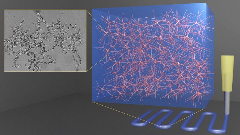
The hydrogel material comes from different-sized seaweed particles. Credit: Orlin Velev, NC State University

Hydrogels merge two physical forms of the same seaweed material for strength, flexibility.

3D-printable gels with improved and highly controlled properties can be created by merging micro- and nano-sized networks of the same materials harnessed from seaweed, according to new research from North Carolina State University. The findings could have applications in biomedical materials — think of biological scaffolds for growing cells — and soft robotics.

Described in the journal Nature Communications, the findings show that these water-based gels — called homocomposite hydrogels — are both strong and flexible. They are composed of alginates — chemical compounds found in seaweed and algae that are commonly used as thickening agents and in wound dressings.

Merging different-size scale networks of the same alginate together eliminates the fragility that can sometimes occur when differing materials are merged together in a hydrogel, says Orlin Velev, S. Frank and Doris Culberson Distinguished Professor of Chemical and Biomolecular Engineering at NC State and corresponding author of the paper.

“Water-based materials can be soft and brittle,” he said. “But these homocomposite materials — soft fibrillar alginate particles inside a medium of alginate — are really two hydrogels in one: one is a particle hydrogel and one is a molecular hydrogel. Merged together they produce a jelly-like material that is better than the sum of its parts, and whose properties can be tuned precisely for shaping through a 3D printer for on-demand manufacturing.”

“We are reinforcing a hydrogel material with the same material, which is remarkable because it uses just one material to improve the overall mechanical properties,” said Lilian Hsiao, an assistant professor of chemical and molecular engineering at NC State and a co-author of the paper. “Alginates are used in wound dressings, so this material potentially could be used as a strengthened 3D-printed bandage or as a patch for wound healing or drug delivery.”

“These types of materials have the potential to be most useful in medical products, in food products as a thickening agent, or in soft robotics,” said Austin Williams, one of the paper’s first coauthors and a graduate student in Velev’s lab.

Future work will attempt to fine-tune this method of merging of homocomposite materials to advance 3D printing for biomedical applications or biomedical injection materials, Velev said.

“This technique may have uses with other types of gels, like those used in coatings or in consumer products,” Hsiao said.

Reference: “Printable homocomposite hydrogels with synergistically reinforced molecular-colloidal networks” by Austin Williams, Sangchul Roh, Alan Jacob, Lilian Hsiao, Orlin D. Velev and Simeon Stoyanov, 14 May 2021, Nature Communications.
DOI: 10.1038/s41467-021-23098-9

Former NC State Ph.D. student Sangchul Roh is the paper’s other first coauthor. Coauthor Simeon Stoyanov from Wageningen University participated in the conception of the new material.

The research is funded by the National Science Foundation under grants CMMI-1825476, CBET-1804462 and ECCS-2025064.

Watch NASA's Ingenuity Mars Helicopter Fly in 3D
Watch NASA’s Ingenuity Mars Helicopter Fly in 3D

A new video of the helicopter’s third flight gives viewers the sensation of standing on the Red Planet and seeing the action firsthand.

When NASA’s Ingenuity Mars Helicopter took to the Martian skies on its third flight on April 25, the agency’s Perseverance rover was there to capture the historic moment. Now NASA engineers have rendered the flight in 3D, lending dramatic depth to the flight as the helicopter ascends, hovers, then zooms laterally off-screen before returning for a pinpoint landing. Seeing the sequence is a bit like standing on the Martian surface next to Perseverance and watching the flight firsthand.

Located on the rover’s mast, or “head,” the zoomable dual-camera Mastcam-Z imager provided the view. Along with producing images that enable the public to follow the rover’s daily discoveries, the cameras provide key data to help engineers navigate and scientists choose interesting rocks to study.

Ingenuity Flies in 3D: After the Mastcam-Z imager aboard NASA’s Perseverance rover captured Ingenuity’s third flight on April 25, the frames of the video that was stitched together were then reprojected to optimize viewing in an anaglyph, or an image seen in 3D when seen through color-filtered glasses. Credit: NASA/JPL-Caltech/ASU/MSSS

Justin Maki, an imaging scientist at NASA’s Jet Propulsion Laboratory in Southern California, led the team that stitched the images into a video. The frames of the video were reprojected to optimize viewing in an anaglyph, or an image seen in 3D when viewed with color-filtered glasses (you can create your own 3D glasses in a few minutes).

Maki’s been creating 3D imaging of Mars since his days as a graduate student processing images from NASA’s Sojourner, the first Mars rover in 1997. But this is the first time he’s created actual 3D video of an aircraft flying on Mars. “The Mastcam-Z video capability was inherited from the Mars Science Laboratory MARDI (MArs Descent Imager) camera,” Maki said. “To be reusing this capability on a new mission by acquiring 3D video of a helicopter flying above the surface of Mars is just spectacular.” The videos of the helicopter are the most extensive 3D video yet from the Mastcam-Z team.

The rover’s drivers and robotic-arm operators use a more sophisticated 3D system to understand exactly how things are positioned on Mars before planning the rover’s movements. But, according to Maki, team members have also been viewing still 3D images for rover-drive planning. “A helicopter flying on Mars opens a new era for Mars exploration. It’s a great demonstration of a new technology for exploration,” he added. “With each flight we open up more possibilities.”

Perseverance Rover’s Mastcam-Z Captures Ingenuity’s Third Flight: NASA’s Ingenuity Mars Helicopter takes off and lands in this video captured on April 25, 2021, by Mastcam-Z, an imager aboard NASA’s Perseverance Mars rover. As expected, the helicopter flew out of its field of vision while completing a flight plan that took it 164 feet (50 meters) downrange of the landing spot. Keep watching, the helicopter will return to stick the landing. Credits: NASA/JPL-Caltech/ASU/MSSS

The April 25 flight brought with it several other firsts, with Ingenuity rising 16 feet (5 meters), then flying downrange 164 feet (50 meters). That was a record until Ingenuity traveled 873 feet (266 meters) on its fourth flight, on April 30. For its fifth flight, on May 7, Ingenuity completed its first one-way trip, traveling 423 feet (129 meters), then reaching an altitude of 33 feet (10 meters) above its new landing field.

The flights began as a technology demonstration intended to prove that powered, controlled flight on Mars is possible. Now they will serve as an operations demonstration, exploring how aerial scouting and other functions could benefit future exploration of Mars.

Probiotic Supplement Use Associated With Fewer Respiratory Symptoms in Overweight and Older People

Probiotic Supplements

Findings provide further evidence of relationship between the gut and lungs.

Daily probiotic use was associated with fewer upper respiratory symptoms in overweight and older people, according to a study that suggests a potential role for probiotics in preventing respiratory infections. The study was selected for presentation at Digestive Disease Week® (DDW) 2021.

“This is not necessarily the most intuitive idea, that putting bacteria into your gut might reduce your risk of respiratory infection,” said Benjamin Mullish, MD, a lead researcher on the study and clinical lecturer in the Division of Digestive Diseases, Imperial College London, England, “but it’s further evidence that the gut microbiome has a complex relationship with our various organ systems. It doesn’t just affect how our gut works or how our liver works, it affects aspects of how our whole body works.”

Researchers re-analyzed detailed daily diaries of 220 patients who participated in an earlier double-blind placebo-controlled study on probiotics and weight loss. Reviewing the entries for common symptoms of upper respiratory infection, including cough, sore throat, and wheezing, researchers found that participants who took probiotics during the six-month study had a 27 percent lower overall incidence of upper respiratory tract symptoms compared to the placebo group. The effect was largest among participants who were aged 45 years or older, as well as those with obesity.

People with obesity are at higher risk for respiratory infections. Previous research has shown that probiotics reduce upper respiratory infections in healthy adults and children, but little data exists on this vulnerable population of older, overweight and people with obesity.

“These findings add to growing interest in the gut-lung axis — how the gut and the lungs communicate with each other,” Dr. Mullish said. “It’s not just the gut sending out signals that affect how the lungs work. It works in both directions. It adds to the story that changes in the gut microbiome can affect large aspects of our health.”

The researchers did not measure immune response, only respiratory symptoms. Future randomized clinical trials could help identify the mechanisms related to the reduction in respiratory symptoms and explore the possible impact of probiotics on the immune system, Dr. Mullish said.

DDW Presentation Details

Dr. Mullish will present data from the study, “Daily probiotic use is associated with a reduced rate of upper respiratory tract symptoms in overweight and obese people,” abstract 739, on Sunday, May 23, at 1:16 p.m. EDT.

Digestive Disease Week® (DDW) is the largest international gathering of physicians, researchers and academics in the fields of gastroenterology, hepatology, endoscopy and gastrointestinal surgery. Jointly sponsored by the American Association for the Study of Liver Diseases (AASLD), the American Gastroenterological Association (AGA) Institute, the American Society for Gastrointestinal Endoscopy (ASGE) and the Society for Surgery of the Alimentary Tract (SSAT), DDW is a fully virtual meeting from May 21-23, 2021. The meeting showcases more than 2,000 abstracts and hundreds of lectures on the latest advances in GI research, medicine and technology.

Hidden Processes Revealed at Work in the Hearts of Massive Stars
3-Solar-Mass Star

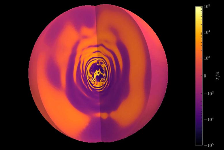
A simulation of a 3-solar-mass star shows the central, convective core and the waves it generates in the rest of the star’s interior. Credit: Philipp Edelmann

Astronomers commonly refer to massive stars as the chemical factories of the Universe. They generally end their lives in spectacular supernovae, events that forge many of the elements on the periodic table. How elemental nuclei mix within these enormous stars has a major impact on our understanding of their evolution prior to their explosion. It also represents the largest uncertainty for scientists studying their structure and evolution.

A team of astronomers led by May Gade Pedersen, a postdoctoral scholar at UC Santa Barbara’s Kavli Institute for Theoretical Physics, have now measured the internal mixing within an ensemble of these stars using observations of waves from their deep interiors. While scientists have used this technique before, this paper marks the first time this has been accomplished for such a large group of stars at once. The results, published in Nature Astronomy, show that the internal mixing is very diverse, with no clear dependence on a star’s mass or age.

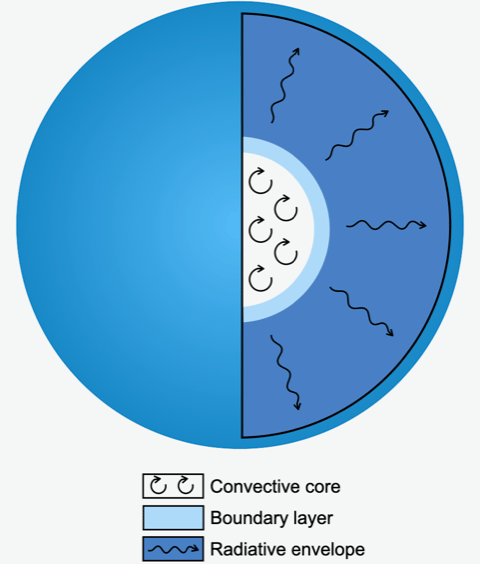
Stars spend the majority of their lives fusing hydrogen into helium deep in their cores. However, the fusion in particularly massive stars is so concentrated at the center that it leads to a turbulent convective core similar to a pot of boiling water. Convection, along with other processes like rotation, effectively removes helium ash from the core and replaces it with hydrogen from the envelope. This enables the stars to live much longer than otherwise predicted.

Astronomers believe this mixing arises from various physical phenomena, like internal rotation and internal seismic waves in the plasma excited by the convecting core. However, the theory has remained largely unconstrained by observations as it occurs so deep within the star. That said, there is an indirect method of peering into stars: asteroseismology, the study and interpretation of stellar oscillations. The technique has parallels to how seismologists use earthquakes to probe the interior of the Earth.

Massive Star Interior

Mixing transports fused material away and replaces it with more hydrogen fuel from the star’s outer layers. Credit: May Gade Pedersen

“The study of stellar oscillations challenges our understanding of stellar structure and evolution,” Pedersen said. “They allow us to directly probe the stellar interiors and make comparisons to the predictions from our stellar models.”

Pedersen and her collaborators from KU Leuven, the University of Hasselt, and the University of Newcastle have been able to derive the internal mixing for an ensemble of such stars using asteroseismology. This is the first time such a feat has been achieved, and was possible thanks only to a new sample of 26 slowly pulsating B-type stars with identified stellar oscillations from NASA’s Kepler mission.

Slowly pulsating B-type stars are between three and eight times more massive than the Sun. They expand and contract on time scales of the order of 12 hours to 5 days, and can change in brightness by up to 5%. Their oscillation modes are particularly sensitive to the conditions near the core, Pedersen explained.

“The internal mixing inside stars has now been measured observationally and turns out to be diverse in our sample, with some stars having almost no mixing while others reveal levels a million times higher,” Pedersen said. The diversity turns out to be unrelated to the mass or age of the star. Rather, it’s primarily influenced by the internal rotation, though that is not the only factor at play.

“These asteroseismic results finally allow astronomers to improve the theory of internal mixing of massive stars, which has so far remained uncalibrated by observations coming straight from their deep interiors,” she added.

The precision at which astronomers can measure stellar oscillations depends directly on how long a star is observed. Increasing the time from one night to one year results in a thousand-fold increase in the measured precision of oscillation frequencies.

“May and her collaborators have really shown the value of asteroseismic observations as probes of the deep interiors of stars in a new and profound way,” said KITP Director Lars Bildsten, the Gluck Professor of Theoretical Physics. “I am excited to see what she finds next.”

The best data currently available for this comes from the Kepler space mission, which observed the same patch of the sky for four continuous years. The slowly pulsating B-type stars were the highest mass pulsating stars that the telescope observed. While most of these are slightly too small to go supernova, they do share the same internal structure as the more massive stellar chemical factories. Pedersen hopes insights gleaned from studying the B type stars will shed light on the inner workings of their higher mass, O type counterparts.

She plans to use data from NASA’s Transiting Exoplanet Survey Satellite (TESS) to study groups of oscillating high-mass stars in OB associations. These groups comprise 10 to more than 100 massive stars between 3 and 120 solar masses. Stars in OB associations are born from the same molecular cloud and share similar ages, she explained. The large sample of stars, and constraint from their common ages, provides exciting new opportunities to study the internal mixing properties of high-mass stars.

In addition to unveiling the processes hidden within stellar interiors, research on stellar oscillations can also provide information on other properties of the stars.

“The stellar oscillations not only allow us to study the internal mixing and rotation of the stars, but also determine other stellar properties such as mass and age,” Pedersen explained. “While these are both two of the most fundamental stellar parameters, they are also some of the most difficult to measure.”

Reference: “Internal mixing of rotating stars inferred from dipole gravity modes” by May G. Pedersen, Conny Aerts, Péter I. Pápics, Mathias Michielsen, Sarah Gebruers, Tamara M. Rogers, Geert Molenberghs, Siemen Burssens, Stefano Garcia and Dominic M. Bowman, 10 May 2021, Nature Astronomy.
DOI: 10.1038/s41550-021-01351-x

Revolutionary Eco-Friendly Plastic: The Future Looks Bright for Infinitely Recyclable Plastic
Plastic Scrap

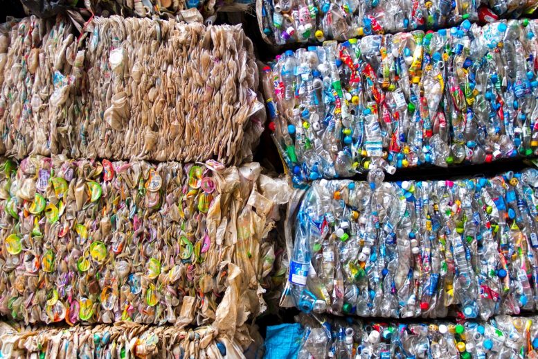
Only about 2% of plastics are fully recycled currently. PDK plastics could solve the single-use crisis.

A new environmental and technological analysis suggests that a revolutionary eco-friendly plastic is almost ready to hit the shelves.

Plastics are a part of nearly every product we use on a daily basis. The average person in the U.S. generates about 100 kg of plastic waste per year, most of which goes straight to a landfill. A team led by Corinne Scown, Brett Helms, Jay Keasling, and Kristin Persson at Lawrence Berkeley National Laboratory (Berkeley Lab) set out to change that.

Less than two years ago, Helms announced the invention of a new plastic that could tackle the waste crisis head on. Called poly(diketoenamine), or PDK, the material has all the convenient properties of traditional plastics while avoiding the environmental pitfalls, because unlike traditional plastics, PDKs can be recycled indefinitely with no loss in quality.

Now, the team has released a study that shows what can be accomplished if manufacturers began using PDKs on a large scale. The bottom line? PDK-based plastic could quickly become commercially competitive with conventional plastics, and the products will get less expensive and more sustainable as time goes on.

“Plastics were never designed to be recycled. The need to do so was recognized long afterward,” explained Nemi Vora, first author on the report and a former postdoctoral fellow who worked with senior author Corinne Scown. “But driving sustainability is the heart of this project. PDKs were designed to be recycled from the get-go, and since the beginning, the team has been working to refine the production and recycling processes for PDK so that the material could be inexpensive and easy enough to be deployed at commercial scales in anything from packaging to cars.”

The study presents a simulation for a 20,000-metric-ton-per-year facility that puts out new PDKs and takes in used PDK waste for recycling. The authors calculated the chemical inputs and technology needed, as well as the costs and greenhouse gas emissions, then compared their findings to the equivalent figures for production of conventional plastics.

“These days, there is a huge push for adopting circular economy practices in the industry. Everyone is trying to recycle whatever they’re putting out in the market,” said Vora. “We started talking to industry about deploying 100% infinitely recycled plastics and have received a lot of interest.”

“The questions are how much it will cost, what the impact on energy use and emissions will be, and how to get there from where we are today,” added Helms, a staff scientist at Berkeley Lab’s Molecular Foundry. “The next phase of our collaboration is to answer these questions.”

Checking the boxes of cheap and easy

To date, more than 8.3 billion metric tons of plastic material have been produced, and the vast majority of this has ended up in landfills or waste incineration plants. A small proportion of plastics are sent to be recycled “mechanically,” meaning they are melted down and then re-shaped into new products. However, this technique has limited benefit. Plastic resin itself is made of many identical molecules (called monomers) bound together into long chains (called polymers). Yet to give plastic its many textures, colors, and capabilities, additives like pigments, heat stabilizers, and flame retardants are added to the resin. When many plastics are melted down together, the polymers become mixed with a slew of potentially incompatible additives, resulting in a new material with much lower quality than newly produced virgin resin from raw materials. As such, less than 10% of plastic is mechanically recycled more than once, and recycled plastic usually also contains virgin resin to make up for the dip in quality.

PDK Plastic Readily Breaks Down

A GIF showing how PDK plastic readily breaks down when put in an acidic solution. The acid helps to break the bonds between the monomers and separate them from the chemical additives that give plastic its look and feel. Credit: Peter Christensen/Berkeley Lab

PDK plastics sidestep this problem entirely – the resin polymers are engineered to easily break down into individual monomers when mixed with an acid. The monomers can then be separated from any additives and gathered to make new plastics without any loss of quality. The team’s earlier research shows that this “chemical recycling” process is light on energy and carbon dioxide emissions, and it can be repeated indefinitely, creating a completely circular material lifecycle where there is currently a one-way ticket to waste.

Yet despite these incredible properties, to truly beat plastics at their own game, PDKs also need to be convenient. Recycling traditional petroleum-based plastic might be hard, but making new plastic is very easy.

“We’re talking about materials that are basically not recycled,” said Scown. “So, in terms of appealing to manufacturers, PDKs aren’t competing with recycled plastic – they have to compete with virgin resin. And we were really pleased to see how cheap and how efficient it will be to recycle the material.”

Scown, who is a staff scientist in Berkeley Lab’s Energy Technologies and Biosciences Areas, specializes in modeling future environmental and financial impacts of emerging technologies. Scown and her team have been working on the PDK project since the outset, helping Helms’ group of chemists and fabrication scientists to choose the raw materials, solvents, equipment, and techniques that will lead to the most affordable and eco-friendly product.

“We’re taking early stage technology and designing what it would look like at commercial-scale operations” using different inputs and technology, she said. This unique, collaborative modeling process allows Berkeley Lab scientists to identify potential scale-up challenges and make process improvements without costly cycles of trial and error.

The team’s report, published in Science Advances, models a commercial-scale PDK production and recycling pipeline based on the plastic’s current state of development. “And the main takeaways were that, once you’ve produced the PDK initially and you’ve got it in the system, the cost and the greenhouse gas emissions associated with continuing to recycle it back to monomers and make new products could be lower than, or at least on par with, many conventional polymers,” said Scown.

Planning to launch

Thanks to optimization from process modeling, recycled PDKs are already drawing interest from companies needing to source plastic. Always looking to the future, Helms and his colleagues have been conducting market research and meeting with people from industry since the project’s early days. Their legwork shows that the best initial application for PDKs are markets where the manufacturer will receive their product back at the end of its lifespan, such as the automobile industry (through trade-ins and take-backs) and consumer electronics (through e-waste programs). These companies will then be able to reap the benefits of 100% recyclable PDKs in their product: sustainable branding and long-term savings.

Workers Sorting Plastic Waste

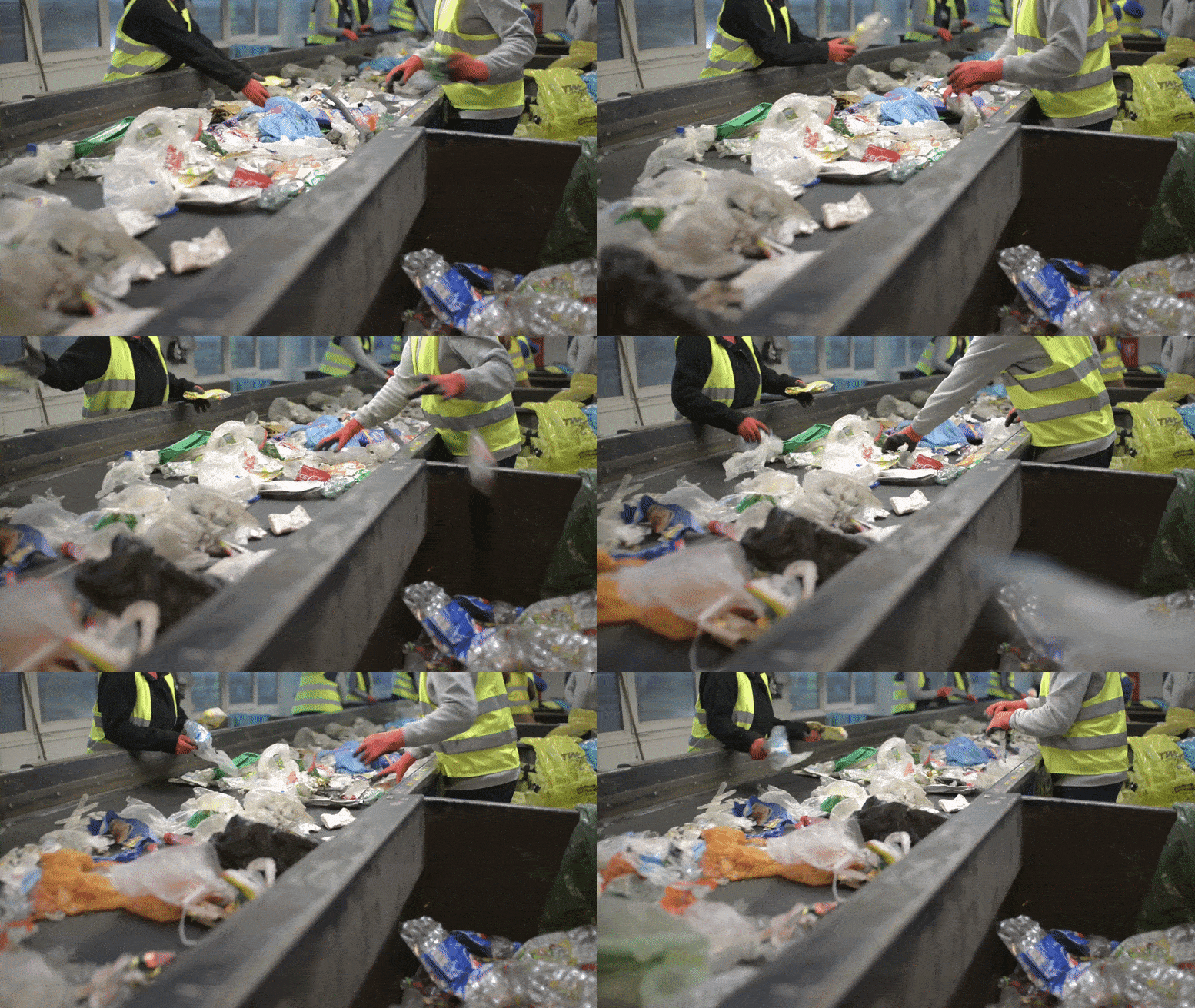
Workers sorting plastic waste.

“With PDKs, now people in industry have a choice,” said Helms. “We’re bringing in partners who are building circularity into their product lines and manufacturing capabilities, and giving them an option that is in line with future best practices.”

Added Scown: “We know there’s interest at that level. Some countries have plans to charge hefty fees on plastic products that rely on non-recycled material. That shift will provide a strong financial incentive to move away from utilizing virgin resins and should drive a lot of demand for recycled plastics.”

After infiltrating the market for durable products like cars and electronics, the team hopes to expand PDKs into shorter-lived, single-use goods such as packaging.

A full-circle future

As they forge plans for a commercial launch, the scientists are also continuing their techno-economic collaboration on the PDK production process. Although the cost of recycled PDK is already projected to be competitively low, the scientists are working on additional refinements to lower the cost of virgin PDK, so that companies are not deterred by the initial investment price.

And true to form, the scientists are working two steps ahead at the same time. Scown, who is also vice president for Life-cycle, Economics & Agronomy at the Joint BioEnergy Institute (JBEI), and Helms are collaborating with Jay Keasling, a leading synthetic biologist at Berkeley Lab and UC Berkeley and CEO of JBEI, to design a process for producing PDK polymers using microbe-made precursor ingredients. The process currently uses industrial chemicals, but was initially designed with Keasling’s microbes in mind, thanks to a serendipitous cross-disciplinary seminar.

“Shortly before we started the PDK project, I was in a seminar where Jay was describing all the molecules that they could make at JBEI with their engineered microbes,” said Helms. “And I got very excited because I saw that some of those molecules were things that we put in PDKs. Jay and I had a few chats, and we realized that nearly the entire polymer could be made using plant material fermented by engineered microbes.”

“In the future, we’re going to bring in that biological component, meaning that we can begin to understand the impacts of transitioning from conventional feedstocks to unique and possibly advantaged bio-based feedstocks that might be more sustainable long term on the basis of energy, carbon, or water intensity of production and recycling,” Helms continued.

“So, where we are now, this is the first step of many, and I think we have a really long runway in front of us, which is exciting.”

Reference: “Leveling the cost and carbon footprint of circular polymers that are chemically recycled to monomer” by Nemi Vora, Peter R. Christensen, Jérémy Demarteau, Nawa Raj Baral, Jay D. Keasling, Brett A. Helms and Corinne D. Scown, 9 April 2021, Science Advances.
DOI: 10.1126/sciadv.abf0187

The Molecular Foundry is a Department of Energy (DOE) Office of Science user facility that specializes in nanoscale science. JBEI is a Bioenergy Research Center funded by DOE’s Office of Science.

This work was supported by the DOE’s Bioenergy Technologies Office and Berkeley Lab’s Laboratory Directed Research and Development (LDRD) program.

The First Frost Is the Deepest – New Discovery May Help Us Grow Crops in Fluctuating Climate
Frost on Arabidopsis thaliana

Frost on Arabidopsis thaliana — new discovery may help us grow crops in fluctuating climate. Credit: John Innes Centre

The first frost of autumn may be grim for gardeners but the latest evidence reveals it is a profound event in the life of plants.

The discovery may affect how we grow crops in a fluctuating climate and help us better understand molecular mechanisms in animals and humans.

Much of our understanding of how plants register temperature at a molecular level has been gained from the study of vernalization — the exposure to an extended period of cold as a preparation for flowering in spring.

Experiments using the model plant Arabidopsis have shown how this prolonged period of cold lifts the brake on flowering, a gene called FLC. This biochemical brake also involves another molecule COOLAIR which is antisense to FLC. This means it lies on the other strand of DNA to FLC and it can bind to FLC and influence its activity.

But less is known about how natural temperature changes affect this process. How does COOLAIR facilitate the shutdown of FLC in nature?

To find out, researchers from the John Innes Centre used naturally occurring types of Arabidopsis grown in different climates.

They measured how much COOLAIR is turned on in three different field sites with varying winter conditions, one in Norwich, UK, one in south Sweden, and one in subarctic northern Sweden.

COOLAIR levels varied among different accessions and different locations. However, researchers spotted something that all the plants had in common — the first time the temperature dropped below freezing there was a peak in COOLAIR.

To confirm this boosting of COOLAIR after freezing they did experiments in temperature-controlled chambers which simulated the temperature changes seen in natural conditions.

They found COOLAIR expression levels rose within an hour of freezing and peaked about eight hours afterwards. There was a small reduction in FLC levels immediately after freezing too, reflecting the relationship between the two key molecular components.

Next, they found a mutant Arabidopsis which produces higher levels of COOLAIR all the time even when it is not cold, and low levels of FLC. When they edited the gene to switch off COOLAIR they found that FLC was no longer suppressed, providing further evidence of this elegant molecular mechanism.

Dr Yusheng Zhao, co-first author of the study said: “Our study shows a new aspect of temperature sensing in plants in natural field conditions. The first seasonal frost serves as an important indicator in autumn for winter arrival. The initial freezing dependent induction of COOLAIR appears to be an evolutionarily conserved feature in Arabidopsis and helps to explain how plants sense environmental signals to begin silencing of the major floral repressor FLC to align flowering with spring.”

The study offers insight into the plasticity in the molecular process of how plants sense temperatures which may help plants adapt to different climates.

Professor Dame Caroline Dean, corresponding author of the study explained: “From the plant’s point of view it gives you a tunable way of shutting off FLC. Any modulation of antisense will switch off sense and from an evolutionary perspective, depending on how efficiently or how fast this happens, and how many cells it happens in, you then have a way of dialing the brake up and down among cells.”

The findings will be helpful for understanding how plants and other organisms sense fluctuating environmental signals and could be translatable to improving crops at a time of climate change.

The discovery will also likely be widely relevant for environmental regulation of gene expression in many organisms because antisense transcription has been shown to alter transcription in yeast and human cells.

Reference: “Natural temperature fluctuations promote COOLAIR regulation of FLC” by Yusheng Zhao1, Pan Zhu1, Jo Hepworth, Rebecca Bloomer, Rea Laila Antoniou-Kourounioti, Jade Doughty, Amelie Heckmann, Congyao Xu, Hongchun Yang and Caroline Dean, 13 May 2021, Genes & Development.
DOI: 10.1101/gad.348362.121

Preventing the Next Pandemic: Scientists Say We Must Regulate Air Like Food and Water

Air Ducts

Humans in the 21st century spend most of their time indoors, but the air we breathe inside buildings is not regulated to the same degree as the food we eat and the water we drink. A group of 39 researchers from 14 countries, including two from the University of Colorado Boulder, say that needs to change to reduce disease transmission and prevent the next pandemic.

In a Perspectives piece published in Science on May 14, 2021, they call for a “paradigm shift” in combating airborne pathogens such as SARS-CoV-2, the virus that causes COVID-19, demanding universal recognition that respiratory infections can be prevented by improving indoor ventilation systems.

“Air can contain viruses just as water and surfaces do,” said co-author Shelly Miller, professor of mechanical and environmental engineering. “We need to understand that it’s a problem and that we need to have, in our toolkit, approaches to mitigating risk and reducing the possible exposures that could happen from build-up of viruses in indoor air.”

The paper comes less than two weeks after the World Health Organization (WHO) changed its website to acknowledge that SARS-CoV-2 is spread predominantly through the air, and 10 months after the WHO acknowledged the potential for aerosol transmission and 239 scientists (including Miller and Jose-Luis Jimenez) signed an open letter to medical communities and governing bodies about the potential risk of airborne transmission. The researchers now call on the WHO and other governing bodies in this new article to extend its indoor air quality guidelines to include airborne pathogens and to recognize the need to control hazards of airborne transmission of respiratory infections.

Ducted Air Pipes

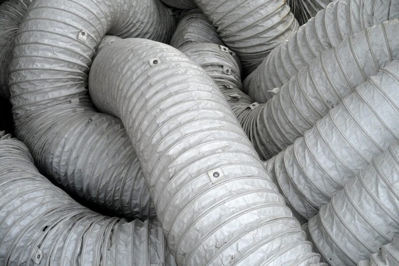
A tangle of ducted air pipes are connected to a portable air unit being used to air condition a large hall. Credit: Martin Visser, Unsplash

Such a shift in ventilation standards should be similar in scale to the 19th century transformation that took place when cities started organizing clean water supplies and centralized sewage systems. But it would also correct a major scientific misperception that arose around the same time.

When people in London were dying of cholera in the 1850s, scientists assumed the disease was airborne. But British physician John Snow discovered that microorganisms in contaminated water were the reason. Similarly, Hungarian physician Ignaz Semmelweis showed that handwashing before delivering a baby greatly reduced postpartum infections. While these discoveries encountered great resistance in their time, scientists eventually agreed that in these cases, water and hands — not air — were the vector for disease.

Then in the early 20th century, American public health expert Charles Chapin erroneously attributed respiratory infections caught in close proximity to other people to large droplets produced by an infected person, which fall quickly to the ground. As a result, he stated that airborne transmission was almost impossible.

Yet in 1945, scientist William Wells published a paper in the predecessor to Science, lamenting that while we were investing in disinfecting water and keeping our food clean, we had done nothing for our indoor air, given the denial of airborne transmission. His research on measles and tuberculosis — caused by airborne pathogens — challenged this notion in the 20th century, but didn’t break it.

Now that the research on SARS-CoV-2 finally has brought to light that many respiratory diseases can be transmitted through the air, researchers argue that we must take action.

“Let’s now not waste time until the next pandemic,” said co-author Jose-Luis Jimenez, fellow in the Cooperative Institute of Research Sciences (CIRES) and professor of chemistry at CU Boulder. “We need a societal effort. When we design a building, we shouldn’t just put in the minimum amount of ventilation that’s possible, but instead we should keep ongoing respiratory diseases, such as the flu, and future pandemics in mind.”

The long-standing misunderstanding of the importance of airborne transmission of pathogens has left a large gap of information in how to best construct and manage building ventilation systems to mitigate the spread of disease — with the exception of some manufacturing, research and medical facilities. Instead, buildings have focused on temperature, odor control, energy use, and perceived air quality. So while there are safety guidelines for chemicals such as carbon monoxide, there are currently no guidelines, globally or in the U.S., that regulate or provide standards for mitigating bacteria or viruses in indoor air resulting from human activities.

“Air in buildings is shared air — it’s not a private good, it’s a public good. And we need to start treating it like that,” said Miller.

Lidia Morawska, lead author on the article and director of Queensland University of Technology’s International Laboratory for Air Quality and Health, said there needs to be a shift away from the perception that we cannot afford the cost of control. She notes that the global monthly cost from COVID-19 had been conservatively estimated as $1 trillion and the cost of influenza in the U.S. alone exceeded $11.2 billion annually.

While detailed economic analysis has yet to be done, estimates suggest necessary investments in building systems may be less than 1% of the construction cost of a typical building.

Ventilation systems should also be demand-controlled to adjust for different room occupancies, and differing activities and breathing rates, such as exercising in a gym versus sitting in a movie theatre, according to Morawska. For spaces that cannot improve ventilation to an appropriate level for the use of the space, she said air filtration and disinfection will be needed.

Because buildings consume over one-third of energy globally, much from heating or cooling outdoor air as it is brought indoors, it would be useful to design a “pandemic mode,” that would allow for buildings to only use more energy when necessary, said Jimenez.

The researchers also call for national comprehensive indoor air quality (IAQ) standards to be developed and enforced by all countries, and for this information to be available to the public.

For this to happen, however, many more than scientists will need to understand its importance.

“I think there is a certain amount of demand that needs to start coming from the consumer and from the person who works in these indoor spaces in order to push change,” said Miller.

Reference: 14 May 2021, Science.
DOI: 10.1126/science.abg2025

Accidental Release From a Lab or Zoonotic Spillover? Scientists Call for Investigation Into the Origins of COVID-19

Virus Lab

Letter From Scientists: Investigate the Origins of COVID-19

More investigation is needed to determine the origin of the COVID-19 pandemic, say Jesse Bloom, Alina Chan, Ralph Baric, David Relman and colleagues in this Letter.

“Theories of accidental release from a lab and zoonotic spillover both remain viable,” they say. “Knowing how COVID-19 emerged is critical for informing global strategies to mitigate the risk of future outbreaks.”

The authors highlight a joint China-World Health Organization (WHO) report into the origins of SARS-CoV-2, some results of which were released in November 2020. “WHO Director-General Tedros Ghebreyesus commented that the report’s consideration of evidence supporting a laboratory accident was insufficient,” they note. “As scientists with relevant expertise, we agree with the WHO director-general, the United States and 13 other countries, and the European Union that greater clarity about the origins of this pandemic is necessary and feasible to achieve,” they say.

They call for an investigation that is “transparent, objective, data-driven, inclusive of broad expertise, subject to independent oversight, and responsibly managed.” In concluding their Letter, they write: “in this time of unfortunate anti-Asian sentiment in some countries, we note that at the beginning of the pandemic, it was Chinese doctors, scientists, journalists, and citizens who shared with the world crucial information about the spread of the virus — often at great personal cost. We should show the same determination in promoting a dispassionate science-based discourse on this difficult but important issue.”

Reference: “Investigate the origins of COVID-19” by J.D. Bloom at Fred Hutchinson Cancer Research Center in Seattle, WA; J.D. Bloom; A. Iwasaki; R. Medzhitov at Howard Hughes Medical Institute in Chevy Chase, MD; Y. Alina Chan; B.E. Deverman at Broad Institute of MIT and Harvard in Cambridge, MA; R.S. Baric at University of North Carolina at Chapel Hill in Chapel Hill, NC; P.J. Bjorkman at California Institute of Technology in Pasadena, CA; S. Cobey at University of Chicago in Chicago, IL; D.N. Fisman at University of Toronto in Toronto, ON, Canada; R. Gupta at Cambridge Institute of Therapeutic Immunology & Infectious Disease in Cambridge, UK; A. Iwasaki; R. Medzhitov at Yale University School of Medicine in New Haven, CT; M. Lipsitch at Harvard T. H. Chan School of Public Health in Boston, MA; R.A. Neher; E. van Nimwegen at University of Basel in Basel, Switzerland; R.A. Neher; E. van Nimwegen at Swiss Institute of Bioinformatics in Basel, Switzerland; R. Nielsen at University of California, Berkeley in Berkeley, CA; N. Patterson at Harvard University in Cambridge, MA; T. Stearns; D.A. Relman at Stanford University in Stanford, CA; M. Worobey at University of Arizona in Tucson, AZ; D.A. Relman at Stanford University School of Medicine in Stanford, CA.
DOI: 10.1126/science.abj0016

Evolutionary Biologists Just Discovered How Some Lizards Are Able to Breathe Underwater
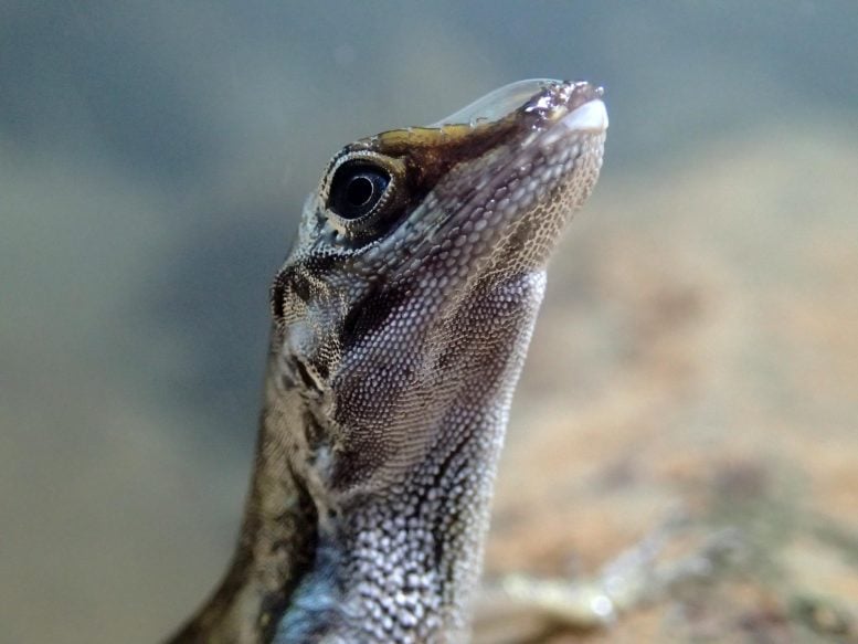
Anolis Lizard With Rebreathing Bubble on Snout

Close-up of an Anolis lizard with a rebreathing bubble on its snout. Credit: Lindsey Swierk

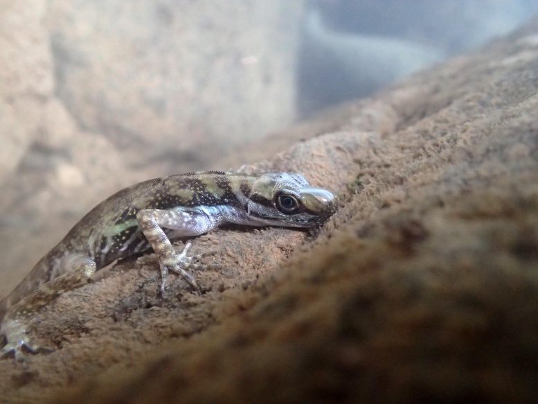
A team of evolutionary biologists from the University of Toronto has shown that Anolis lizards, or anoles, are able to breathe underwater with the aid of a bubble clinging to their snouts.

Anoles are a diverse group of lizards found throughout the tropical Americas. Some anoles are stream specialists, and these semi-aquatic species frequently dive underwater to avoid predators, where they can remain submerged for as long as 18 minutes.

“We found that semi-aquatic anoles exhale air into a bubble that clings to their skin,” says Chris Boccia, a recent Master of Science graduate from the Faculty of Arts & Science’s Department of Ecology & Evolutionary Biology (EEB). Boccia is lead author of a paper describing the finding published this week in Current Biology.

“The lizards then re-inhale the air,” says Boccia, “a maneuver we’ve termed ‘rebreathing’ after the scuba-diving technology.”

The researchers measured the oxygen (O2) content of the air in the bubbles and found that it decreased over time, confirming that rebreathed air is involved in respiration.

Rebreathing likely evolved because the ability to stay submerged longer increases the lizard’s chances of eluding predators.

Submerged Anolis Lizard With Rebreathing Bubble

A submerged Anolis lizard with a rebreathing bubble on its snout. Credit: Lindsey Swierk

The authors studied six species of semi-aquatic anoles and found that all possessed the rebreathing trait, despite most species being distantly related. While rebreathing has been studied extensively in aquatic arthropods like water beetles, it was not expected in lizards because of physiological differences between arthropods and vertebrates.

“Rebreathing had never been considered as a potential natural mechanism for underwater respiration in vertebrates,” says Luke Mahler, an assistant professor in EEB and Boccia’s thesis supervisor. “But our work shows that this is possible and that anoles have deployed this strategy repeatedly in species that use aquatic habitats.”

Mahler and co-author Richard Glor, from the University of Kansas, first observed anoles rebreathing in Haiti in 2009 but were unable to carry out further observations or experiments. Another co-author, Lindsey Swierk, from Binghamton University, State University of New York, described the same behavior in a Costa Rican species in 2019. These early observations suggested that rebreathing was an adaptation for diving, but this idea had not been tested until now.

Boccia became interested in aquatic anoles after encountering one in Panama. He began his rebreathing investigations in Costa Rica in 2017 and continued the research in Colombia and Mexico.

As the authors point out, the rebreathing trait may have developed because anoles’ skin is hydrophobic — it repels water — a characteristic that likely evolved in anoles because it protects them from rain and parasites. Underwater, air bubbles cling to hydrophobic skin, and the ability to exploit these bubbles for breathing developed as a result.

While further work is required to understand how the process works in detail, Boccia, Mahler, and their co-authors suggest different ways in which rebreathing may function.

In its simplest form, the air bubble on a lizard’s snout likely acts like a scuba tank, providing a submerged animal with a supply of air in addition to the air in its lungs. This is what aquatic arthropods like water beetles do to extend the time they can remain submerged.

The researchers also suggest that the rebreathing process may facilitate using air found in a lizard’s nasal passages, mouth, and windpipe that would otherwise not be used by the lizard in breathing.

The bubble may also help rid waste carbon dioxide (CO2) from exhaled air through a process other researchers have already observed in aquatic arthropods. Those studies concluded that because CO2 is highly soluble in water and because the level of CO2 in the bubbles is higher than in the surrounding water, exhaled CO2 dissolves into the surrounding water rather than being rebreathed.

Finally, the authors speculate that the bubble may act as a gill and absorb oxygen from the water — again, something already observed in arthropods. Boccia and Mahler are planning further research to confirm if these rebreathing processes are occurring with anoles.

According to Mahler, “This work enriches our understanding of the creative and unexpected ways that organisms meet the challenges posed by their environments. That is valuable in its own right, but discoveries like this can also be valuable to humans as we seek solutions to our own challenging problems.”

“It’s too early to tell if lizard rebreathing will lead to any particular human innovations,” says Boccia, “But biomimicry of rebreathing may be an interesting proposition for several fields — including scuba-diving rebreathing technology, which motivated our naming of this phenomenon.”

Reference: “Repeated evolution of underwater rebreathing in diving Anolis lizards” by Christopher K. Boccia, Lindsey Swierk, Fernando P. Ayala-Varela, James Boccia, Isabela L. Borges, Camilo Andres Estupiñán, Alexandra M. Martin, Ramón E. Martínez-Grimaldo, Sebastian Ovalle, Shreeram Senthivasan, Ken S. Toyama, María del Rosario Castañeda, Andrés García, Richard E. Glor and D. Luke Mahler, 12 May 2021, Current Biology.
DOI: 10.1016/j.cub.2021.04.040

Mahler’s participation in the research was supported by an NSERC Discovery Grant and a Harvard University Ken Miyata Field Research Award. Boccia’s participation was supported by an NSERC CGS M Grant, a National Geographic Young Explorer Grant and a Sigma Xi Grant in Aid of Research.

How to Cloak an Object to Become Invisible to a Thermal Camera

How to Thermally Cloak an Object

Theoretical method can make objects invisible to a thermal camera, or mimic a different object.

Can you feel the heat? To a thermal camera, which measures infrared radiation, the heat that we can feel is visible, like the heat of a traveler in an airport with a fever or the cold of a leaky window or door in the winter.

In a paper published in Proceedings of the Royal Society A: Mathematical, Physical and Engineering Sciences, an international group of applied mathematicians and physicists, including Fernando Guevara Vasquez and Trent DeGiovanni from the University of Utah, report a theoretical way of mimicking thermal objects or making objects invisible to thermal measurements. And it doesn’t require a Romulan cloaking device or Harry Potter’s invisibility cloak. The research is funded by the National Science Foundation.

The method allows for fine-tuning of heat transfer even in situations where the temperature changes in time, the researchers say. One application could be to isolate a part that generates heat in a circuit (say, a power supply) to keep it from interfering with heat sensitive parts (say, a thermal camera). Another application could be in industrial processes that require accurate temperature control in both time and space, for example controlling the cooling of a material so that it crystallizes in a particular manner.

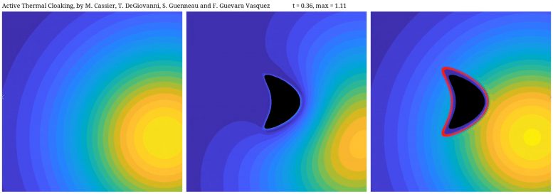
Watch a visualization of how the method cloaks a kite-shaped object here:

Left to right: 1. temperature of a plate subject to a point source firing at time t=0 (this could be e.g. a laser pulse). 2. temperature of the plate with a “kite” object present. As you can see the isotherms, or temperature contours, are deformed by the presence of the object and this can be used by an observer to detect and locate the kite. 3. the kite object is surrounded by our active cloak. Now the isotherms look exactly like the ones in the case where the object is not present, which hides the kite object. Credit: Fernando Guevara Vasquez/University of Utah

Or watch how it works for a Homer Simpson-shaped object here:

Left to right: 1. temperature of a plate subject to point source firing at time t=0 (this could be e.g. a laser pulse). 2. temperature of the plate with an object present. As you can see the isotherms, or temperature contours, are deformed by the presence of the object and this can be used by an observer to detect and locate the object. 3. The object is surrounded by our active cloak. Now the isotherms look exactly like the ones in the case where the object is not present, which hides the object. Credit: Fernando Guevara Vasquez

Cloaking or invisibility devices have long been elements of fictional stories, but in recent years scientists and engineers have explored how to bring science fiction into reality. One approach, using metamaterials, bends light in such a way as to render an object invisible.

Just as our eyes see objects if they emit or reflect light, a thermal camera can see an object if it emits or reflects infrared radiation. In mathematical terms, an object could become invisible to a thermal camera if heat sources placed around it could mimic heat transfer as if the object wasn’t there.

The novelty in the team’s approach is that they use heat pumps rather than specially crafted materials to hide the objects. A simple household example of a heat pump is a refrigerator: to cool groceries it pumps heat from the interior to the exterior. Using heat pumps is much more flexible than using carefully crafted materials, Guevara says. For example, the researchers can make one object or source appear as a completely different object or source. “So at least from the perspective of thermal measurements,” Guevara says, “they can make an apple appear as an orange.”

The researchers carried out the mathematical work needed to show that, with a ring of heat pumps around an object, it’s possible to thermally hide an object or mimic the heat signature of a different object.

The work remains theoretical, Guevara says, and the simulations assume a “probing” point source of heat that would reflect or bend around the object – the thermal equivalent of a flashlight in a dark room.

The temperature of that probing source must be known ahead of time, a drawback of the work. However the approach is within reach of current technology by using small heat pumps called Peltier elements that transport heat by passing an electrical current across a metal-metal junction. Peltier elements are already widely used in consumer and industrial applications.

The researchers envision their work could be used to accurately control the temperature of an object in space and time, which has applications in protecting electronic circuits. The results, the researchers say, could also be applied to accurate drug delivery, since the mathematics of heat transfer and diffusion are similar to those of the transfer and diffusion of medications. And, they add, the mathematics of how light behaves in diffuse media such as fog could lead to applications in visual cloaking as well.

Reference: “Active thermal cloaking and mimicking” by Maxence Cassier, Trent DeGiovanni, Sébastien Guenneau and Fernando Guevara Vasquez, 12 May 2021, Proceedings of the Royal Society A: Mathematical, Physical and Engineering Sciences.
DOI: 10.1098/rspa.2020.0941

In addition to Guevara and DeGiovanni, Maxence Cassier, CNRS Researcher at the Fresnel Institute in Marseille, France and Sébastien Guenneau, CNRS researcher, UMI 2004 Abraham de Moivre-CNRS, Imperial College London, London, UK co-authored the study.

Prehistoric Fossilized Footprints Show Earliest Known Evidence of Mammals at the Seashore
Coryphodon Reconstruction

A reconstruction of the brown-bear-sized mammals (Coryphodon) that made thousands of tracks in a 58-million-year-old, brackish water lagoon in what is now southern Wyoming. Credit: Anton Wroblewski

Today, the rocks of the Hanna Formation in south-central Wyoming are hundreds of miles away from the nearest ocean. But around 58 million years ago, Wyoming was oceanfront property, with large hippo-like mammals traipsing through nearshore lagoons.

In a study published in Scientific Reports, geologist Anton Wroblewski, an adjunct associate professor in the Department of Geology and Geophysics, and applied biodiversity scientist Bonnie Gulas-Wroblewski of the Texas A&M Natural Resources Institute, report the discovery of several sets of fossilized tracks, likely from the brown bear-sized Coryphodon, that represent the earliest known evidence of mammals gathering near an ocean.

“Trace fossils like footprints record interactions between organisms and their environments, providing information that body fossils alone cannot,” Wroblewski says. “In this case, trace fossils show that large- bodied mammals were regularly using marine environments only eight million years after non-avian dinosaurs went extinct.”

Anton Wroblewski Points to Mammal Track Underprints

Anton Wroblewski points to an underprint made 58 million years ago by a heavy mammal (likely Coryphodon) walking on the deltaic deposits above. Underprints form when sediment is displaced downward by footsteps from heavy animals.” Credit: Anton Wroblewski

The tracks that the Drs. Wroblewski found in the Hanna Formation of Wyoming include underprints, impressions in soft sediment made when heavy animals walk on overlying sediment layers, as well as prints pressed into the surfaces of ancient tidal flats. Now preserved in sandstone, the tracks are more than half a mile (one kilometer) long and were made by two different animals, one with four toes and one with five. The five-toed tracks are consistent with Coryphodon, a semi-aquatic mammal similar to a hippopotamus. The owner of the four-toed tracks remains a mystery.

Mammal Tracks From Paleocene Lagoon

Section of the 58-million-year-old tracksite demonstrating near-vertical tilting of the originally horizontal bedding with three separate trackways made by five-toed mammals walking in parallel. Credit: Anton Wroblewski

“Paleontologists have been working in this area for thirty years, but they’ve been looking for bones, leaf fossils, and pollen, so they didn’t notice footprints or trackways,” Wroblewski says. He first saw the tracks in September 2019. “When I found them, it was late afternoon and the setting sun hit them at just the right angle to make them visible on the tilted slabs of sandstone. At first, I couldn’t believe what I was seeing; I had walked by this outcrop for years without noticing them. Once I saw the first few, I followed out the ridge of sandstone and realized they were part of a much larger, more extensive trackway.”

Fossilized plants and pollen helped the researchers determine the age of the tracks to be around 58 million years old, during the Paleocene epoch. Before this finding, the earliest known evidence of mammals interacting with marine environments came from the Eocene epoch, around 9.4 million years later. Wroblewski says that the Hanna Formation tracks are the first Paleocene mammal tracks found in the USA and only the fourth in the world, with two sets of tracks previously found in Canada and one in Svalbard, Norway. It’s also the largest accumulation of Paleocene mammal tracks in the world in both aerial extent and the absolute number of tracks, he says. With at least two species leaving the tracks, it’s also the most taxonomically diverse.

Today’s large mammals congregate near marine environments for a variety of reasons, including protection from predators and biting insects, foraging for unique foods, and access to salt sources, which may have been limited in the tropical forests of North America during the Paleocene. The researchers say ancient mammals may have had similar reasons for seeking out a day at the beach.

The research shows, Wroblewski says, that hypotheses of behavior and evolution based on isotopic, molecular and body fossil data can be empirically tested using trace fossils. “No other line of evidence directly records behaviors of extinct organisms preserved in their preferred habitats,” he says. “There’s still a lot of important information out there in the rocks, waiting for somebody to spot it when the lighting is just right!”

Reference: 13 May 2021, Scientific Reports.
DOI: 10.1038/s41598-021-88412-3

Supercomputer Simulations Reveal How Dominant COVID-19 Strain Binds to Host, Succumbs to Antibodies
Dominant G-Form Spike Protein

Supercomputer simulations at Los Alamos National Laboratory demonstrated that the G form of SARS-CoV-2, the dominant strain of the virus causing COVID-19, mutated to a conformation that allows it to more easily attach to host receptors, while also being more susceptible to antibodies than the original D form. Credit: Los Alamos National Laboratory

Dominant G-form Spike protein ‘puts its head up’ more frequently to latch on to receptors, but that makes it more vulnerable to neutralization.

Large-scale supercomputer simulations at the atomic level show that the dominant G form variant of the COVID-19-causing virus is more infectious partly because of its greater ability to readily bind to its target host receptor in the body, compared to other variants. These research results from a Los Alamos National Laboratory–led team illuminate the mechanism of both infection by the G form and antibody resistance against it, which could help in future vaccine development.

“We found that the interactions among the basic building blocks of the Spike protein become more symmetrical in the G form, and that gives it more opportunities to bind to the receptors in the host — in us,” said Gnana Gnanakaran, corresponding author of the paper published recently in Science Advances. “But at the same time, that means antibodies can more easily neutralize it. In essence, the variant puts its head up to bind to the receptor, which gives antibodies the chance to attack it.”

Researchers knew that the variant, also known as D614G, was more infectious and could be neutralized by antibodies, but they didn’t know how. Simulating more than a million individual atoms and requiring about 24 million CPU hours of supercomputer time, the new work provides molecular-level detail about the behavior of this variant’s Spike.

Current vaccines for SARS-CoV-2, the virus that causes COVID-19, are based on the original D614 form of the virus. This new understanding of the G variant — the most extensive supercomputer simulations of the G form at the atomic level — could mean it offers a backbone for future vaccines.

The team discovered the D614G variant in early 2020, as the COVID-19 pandemic caused by the SARS-CoV-2 virus was ramping up. These findings were published in Cell. Scientists had observed a mutation in the Spike protein. (In all variants, it is the Spike protein that gives the virus its characteristic corona.) This D614G mutation, named for the amino acid at position 614 on the SARS-CoV-2 genome that underwent a substitution from aspartic acid, prevailed globally within a matter of weeks.

The Spike proteins bind to a specific receptor found in many of our cells through the Spike’s receptor binding domain, ultimately leading to infection. That binding requires the receptor binding domain to transition structurally from a closed conformation, which cannot bind, to an open conformation, which can.

The simulations in this new research demonstrate that interactions among the building blocks of the Spike are more symmetrical in the new G-form variant than those in the original D-form strain. That symmetry leads to more viral Spikes in the open conformation, so it can more readily infect a person.

A team of postdoctoral fellows from Los Alamos — Rachael A. Mansbach (now assistant professor of Physics at Concordia University), Srirupa Chakraborty, and Kien Nguyen — led the study by running multiple microsecond-scale simulations of the two variants in both conformations of the receptor binding domain to illuminate how the Spike protein interacts with both the host receptor and with the neutralizing antibodies that can help protect the host from infection. The members of the research team also included Bette Korber of Los Alamos National Laboratory, and David C. Montefiori, of Duke Human Vaccine Institute.

The team thanks Paul Weber, head of Institutional Computing at Los Alamos, for providing access to the supercomputers at the Laboratory for this research.

Reference: “The SARS-CoV-2 Spike variant D614G favors an open conformational state” by Rachael A. Mansbach, Srirupa Chakraborty, Kien Nguyen, David C. Montefiori, Bette Korber, S. Gnanakaran, 16 April 2021, Science Advances. 
DOI: 10.1126/sciadv.abf3671

Funding: The project was supported by Los Alamos Laboratory Directed Research and Development project 20200706ER, Director’s Postdoctoral fellowship, and the Center of Nonlinear Studies Postdoctoral Program at Los Alamos.

African Forest Elephants Are Now Critically Endangered – Here's How to Count Them
African Forest Elephants Are Now Critically Endangered – Here’s How to Count Them
Forest Elephant

A team of scientists compared methodologies to count African forest elephants (Loxodonta cyclotis), which were recently acknowledged by IUCN as a separate, Critically Endangered species from African savannah elephants. Credit: WCS Gabon

Study compared different methodologies using dung, DNA analysis, and camera traps.

A team of scientists led by the Wildlife Conservation Society (WCS) and working closely with experts from the Agence Nationale des Parcs Nationaux du Gabon (ANPN) compared methodologies to count African forest elephants (Loxodonta cyclotis), which were recently acknowledged by IUCN as a separate, Critically Endangered species from African savannah elephants. The study is part of a larger initiative in partnership with Vulcan Inc. to provide the first nationwide census in Gabon for more than 30 years. The results of the census are expected later this year.

Contrary to savannah elephants (Loxodonta africana) which can be counted directly, usually through aerial survey, accurately censusing elusive forest elephants is more challenging and refinements of methods were needed. Publishing a new survey method to counting forest elephants in the journal Global Ecology and Conservation, the team compared traditional methodologies to count elephant dung piles along line transects, with spatial capture-recapture (SCR) techniques using both camera traps and DNA dung analysis. SCR estimates populations by measuring how many times and in what location individual animals are recounted.

Said the study’s lead author, Alice Laguardia of WCS’s Gabon Program: “The more accurately we can count forest elephants, the more we can measure whether conservation efforts are successful.  We are hopeful that the results of this study will help governments and conservation partners protect this Critically Endangered species throughout its range.”

Researchers assessed the performance of the methodologies to three relatively large forest elephant populations in Gabon. They found that the SCR method that used DNA sampling of dung was comparable in accuracy to the line transect method but less expensive on larger scales.

Stephanie Bourgeois, coauthor and geneticist at ANPN, said: “Testing of this new DNA approach has been made possible by the recent development of novel genetics techniques by ANPN and the creation of a new genetics lab in Gabon enabling to perform all DNA analyses in-country.”

SCR Camera trap surveys were more precise on smaller scales but more expensive. The authors recommend that the use of both SCR methods, and their development, continue. They say that future findings and improvements should be compiled across studies to ensure their robust evolution as an option for monitoring the African forest elephant across its range and inform strategies and action for its conservation.

Forest elephants have been decimated by ivory poachers in recent years. A WCS-led census released in 2014 documented a 65 percent decline in forest elephant numbers between 2002 and 2013. Through this new study, researchers will gain a better understanding of how many forest elephants remain and where they reside. Efforts have been focused on Gabon as it is thought to harbor more than 50 percent of the remaining forest elephant population, despite accounting for less than 15 percent of the species’ range, making Gabon the most important country for forest elephant conservation. 

“As long as ivory is a precious commodity, elephants will be at risk,” said Lee White, the Gabonese Minister of Water, Forests, the Seas, the Environment, charged with Climate Change & Land Use Planning. “In Africa there is a clear link between environmental governance, peace, and security. Countries that have lost their elephant populations have all too often descended into civil strife. Through the results of this study we hope to obtain a clear picture of the trend of poaching and elephant populations in all of Gabon.”

“Vulcan recognizes the significant role of accurate population data for conservation management and policy decisions,” said Ted Schmitt, director, conservation at Vulcan Inc. “By providing timely census data, we can fill critical knowledge gaps and enable prioritization of conservation resources. We are pleased to be part of this effort with Wildlife Conservation Society and the government of Gabon to help preserve this important species.” 

Reference: “Assessing the feasibility of density estimation methodologies for African forest elephant at large spatial scales” by A. Laguardia, K. S. Gobush, S. Bourgeois, S. Strindberg, G. Abitsi, F. Ebouta, J. M. Fay, A. M. Gopalaswamy, F. Maisels, R. Ogden, L. J. T. White and E. J. Stokes, 26 March 2021, Global Ecology and Conservation.
DOI: 10.1016/j.gecco.2021.e01550

Funding for this critical work was provided by Vulcan Inc., a Seattle company founded by the late philanthropist Paul G. Allen and his sister Jody Allen, who currently serves as chair.

Solving The Mystery of the Pointy Droplets

Credit: Leiden University

A certain type of oil droplets changes shape when cooled and shrunk: from spherical through icosahedral to flat hexagonal. Two competing theories couldn’t fully explain this, but now, a Physical Review Letter by Ireth García-Aguilar and Luca Giomi solves the mystery.

It was an accidental discovery. Bulgarian researchers at Sofia University were studying small oily droplets of alkanes in water, stabilized with soap-like surfactant-molecules. “These are similar to the emulsion droplets in mayonnaise,” says Luca Giomi, “and in addition, they are enclosed in a frozen monolayer of alkane molecules and surfactants.”

When the Bulgarians were playing around with them, they realized that something special was going on. When the temperature was lowered, the droplets shifted from ordinary spherical shapes to odd, crystal-like icosahedral shapes. At even lower temperatures, they morphed into four-sided rhombuses or hexagons, with growing tentacles at the corners.

Around the same time, another group at Bar-Ilan University in Israel led by Eli Sloutskin, a coauthor of this letter, made similar observations and further realized that small droplets were more prone to change their shape compared to large droplets.

Hexagonal Liquid Drops. Credit: N Denkov et al. Nature 1-4 (2015) doi:10.1038/nature16189

Exotic

“This is inspiring, it’s very exotic and something you wouldn’t expect,” says Giomi. Normally, large elastic sheets are floppier and more prone to bending than small sheets. “One can verify this by holding a sheet of paper on one side: an A4 sheet will immediately bend under its own weight, but a smaller sheet, such a post stamp, will remain straight. The larger the sheet, the higher the torque it experiences, the easier it bends.”

The group at Sofia University itself advanced a theory in which a special thin layer below the surfactants layer causes the edges, “but later, detailed microscopy images by Sloutskin’s lab, didn’t see such a layer,” says Giomi.

In order to explain the shape transformations as well as the anomalous size dependence, Leiden physicists had to include in their model four different ingredients: surface tension, gravity, defects, and spontaneous curvature. The latter is an effect of the shape of molecules that form the solid layer. When long molecules are stacked like matches in a box, the interface is flat, but when one of the molecules’ ends is fatter than the other, the resulting membrane may have a preferred curvature. 

Weird tentacles

While defects and gravity tend to bend the droplets, surface tension tends to restore the spherical shape. But, in the presence of spontaneous curvature, this effect becomes weaker as droplets become smaller, thus rendering small droplets prone to faceting. This explains the mysterious behavior, the researchers write in a paper in Physical Review Letters

One thing remains to be explained, however: the weird tentacles that develop at the lowest temperatures. “But we do have ideas,” says Giomi.

This type research is foundational and curiosity-driven, he adds. However, the behavior of living cells is always an inspiration. “Biological cells have an extraordinary capacity of changing their shapes with different circumstances.”

One of Giomi’s research topics is how cancer cells manage to split off their main tumor and migrate within the body to form deadly metastases. Giomi: “Cancer cells have to undergo dramatic shape changes in order to do so.” Understanding how simple micron-sized objects can autonomously adjust their shape may be critical to deciphering these processes.

Reference: “Faceting and Flattening of Emulsion Droplets: A Mechanical Model” by Ireth García-Aguilar, Piermarco Fonda, Eli Sloutskin, and Luca Giomi, 21 January 2021, Physical Review Letters.
DOI: 10.1103/PhysRevLett.126.038001

Signs of a Puzzling State of Matter Discovered in a Superconductor
Untangling Three Exotic States in a High-Temperature Superconductor

Scientists at SLAC National Accelerator Laboratory used an improved X-ray technique to explore exotic states of matter in an unconventional superconductor that conducts electricity with 100% efficiency at relatively high temperatures. They glimpsed the signature of a state known as pair density waves (PDW), and confirmed that it intertwines with another phase known as charge density wave (CDW) stripes – wavelike patterns of higher and lower electron density in the material. CDWs, in turn, are created when spin density waves (SDWs) emerge and intertwine. Credit: Jun-Sik Lee/SLAC National Accelerator Laboratory

Known as “pair-density waves,” it may be key to understanding how superconductivity can exist at relatively high temperatures.

Unconventional superconductors contain a number of exotic phases of matter that are thought to play a role, for better or worse, in their ability to conduct electricity with 100% efficiency at much higher temperatures than scientists had thought possible – although still far short of the temperatures that would allow their wide deployment in perfectly efficient power lines, maglev trains and so on.

Now scientists at the Department of Energy’s SLAC National Accelerator Laboratory have glimpsed the signature of one of those phases, known as pair-density waves or PDW, and confirmed that it’s intertwined with another phase known as charge density wave (CDW) stripes – wavelike patterns of higher and lower electron density in the material.

Observing and understanding PDW and its correlations with other phases may be essential for understanding how superconductivity emerges in these materials, allowing electrons to pair up and travel with no resistance, said Jun-Sik Lee, a SLAC staff scientist who led the research at the lab’s Stanford Synchrotron Radiation Lightsource (SSRL).

Even indirect evidence of the PDW phase intertwined with charge stripes, he said, is an important step on the long road toward understanding the mechanism behind unconventional superconductivity, which has eluded scientists over more than 30 years of research.

Lee added that the method his team used to make this observation, which involved dramatically increasing the sensitivity of a standard X-ray technique known as resonant soft X-ray scattering (RSXS) so it could see the extremely faint signals given off by these phenomena, has potential for directly sighting both the PDW signature and its correlations with other phases in future experiments. That’s what they plan to work on next.

The scientists described their findings in an article published in Physical Review Letters.

Untangling superconductor secrets

The existence of the PDW phase in high-temperature superconductors was proposed more than a decade ago and it’s become an exciting area of research, with theorists developing models to explain how it works and experimentalists searching for it in a variety of materials.

In this study, the researchers went looking for it in a copper oxide, or cuprate, material known as LSCFO for the elements it contains ­– lanthanum, strontium, copper, iron and oxygen. It’s thought to host two other phases that may intertwine with PDW: charge density wave stripes and spin density wave stripes.

The nature and behavior of charge and spin stripes have been explored in a number of studies, but there had been only a few indirect glimpses of PDW – much like identifying an animal from its tracks – and none made with X-ray scattering techniques. Because X-ray scattering reveals the behavior of an entire sample at once, it’s thought to be the most promising way to clarify whether PDW exists and how it relates to other key phases in cuprates, Lee said.

Over the past few years, the SSRL team has worked on increasing the sensitivity of RSXS so it could capture the signals they were looking for.

Postdoctoral researcher Hai Huang and SLAC staff engineer Sang-Jun Lee used the improved technique in this study. They scattered X-rays off LSCFO and into a detector, forming patterns that revealed what was going on inside the material. As they dropped the temperature of the material toward its superconducting range, spin stripes appeared and intertwined to form charge stripes, and those charge stripes were then associated with the emergence of two-dimensional fluctuations that are the hallmark of PDW.

The researchers said these results not only demonstrate the value of the new RSXS approach, but also support the possibility that the PDW is present not just in this material, but in all of the superconducting cuprates.

Reference: “Two-Dimensional Superconducting Fluctuations Associated with Charge-Density-Wave Stripes in La1.87Sr0.13Cu0.99Fe0.01O4” by H. Huang, S.-J. Lee, Y. Ikeda, T. Taniguchi, M. Takahama, C.-C. Kao, M. Fujita, and J.-S. Lee, 21 April 2021, Physical Review Letters.
DOI: 10.1103/PhysRevLett.126.167001

A research team led by Masaki Fujita at Tohoku University in Japan grew the high-quality LSCFO crystal used in the experiment and conducted preliminary tests on it there. The research was funded by the DOE Office of Science. SSRL is a DOE Office of Science user facility.